Search results (53 results)
-
 Sclerochoroidal Calcification
Apr 24 2025 by Virginia Gebhart
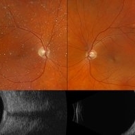
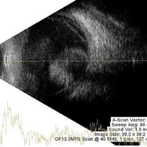
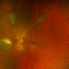
70 year old male referred for amelanotic lesion in the STA OU. Ultrasound shows slightly elevated lesions with hyperreflectivity and posterior shadowing with reduplication artifact consistent with sclerochoroidal calcification. Recommend yearly observation.
Photographer: Virginia Gebhart, Retina Consultants of Carolina
Imaging device: Optos California, Ellex Eye Cubed
Condition/keywords: asteroid hyalosis, B scan ultrasound, sclerochoroidal calcification
-
 Open Funnel (Transversal)
Apr 10 2025 by Gustavo Uriel Fonseca Aguirre
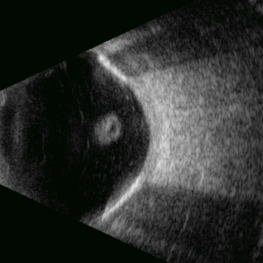
This B-mode transverse ultrasound scan reveals a chronic rhegmatogenous retinal detachment, demonstrating a funnel-shaped configuration with a narrow intraluminal space. Two hyperechoic choroidal calcifications are present, indicative of chronicity.
Photographer: Gustavo U. Fonseca Aguirre, Hospital Conde de Valenciana, Ciudad de México
Condition/keywords: open funnel RD, Retina detachment
-
 Calcification of the Retina
Apr 7 2025 by Gustavo Uriel Fonseca Aguirre
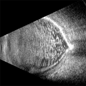
B-mode ultrasound of a vitrectomized eye reveals emulsified silicone oil in the vitreous cavity, retinal detachment, and calcification of the retina and optic nerve head.
Photographer: Gustavo U. Fonseca Aguirre, Hospital Conde de Valenciana, Ciudad de México
Condition/keywords: calcification, Retina detachment, vitrectomy
-
 Optic Nerve Head Drusen With Angiod Streaks in Hyperphosphatemic Familial Tumoral Calcinosis
Aug 8 2024 by Hemanth Murthy, MBBS, MD, FASRS
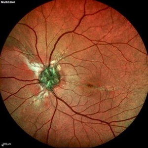
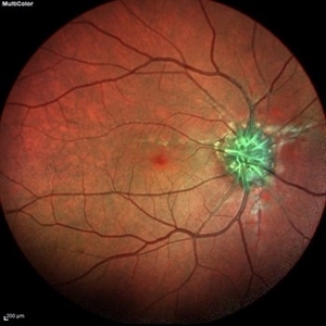
Multicolor image of left eye of 53 year female patient with decreased vision in left eye. Patient gives history of multiple joint swellings with multiple dental procedures due to calcification of the roots. She had type2 MNV demonstrated on OCT and OCTA. Her blood reports showed elevated serum phosphorus (6.4 mg/dl) with normal serum calcium, vitamin D and parathyroid hormone. Her fibroblast growth factor 23 was markedly elevated(>1500RU/ml).
Photographer: Mr Veda Vyas
Condition/keywords: Optic disc drusen and Angiod streaks
-
 Optic Nerve Head Drusen With Angiod Streaks in Hyperphosphatemic Familial Tumoral Calcinosis
Aug 8 2024 by Hemanth Murthy, MBBS, MD, FASRS
Multicolor image of right eye of 53 year female patient with decreased vision in left eye. Patient gives history of multiple joint swellings with multiple dental procedures due to calcification of the roots. She showed type2 MNV on OCT and OCTA. Her blood reports showed elevated serum phosphorus (6.4 mg/dl) with normal serum calcium, vitamin D and parathyroid hormone. Her fibroblast growth factor 23 was markedly elevated(>1500RU/ml).
Photographer: Mr Veda Vyas
Condition/keywords: Optic disc drusen and Angiod streaks
-
 Optic Nerve Head Drusen With Angiod Streaks in Phosphatemic Familial Tumoral Calcinosis
Aug 8 2024 by Hemanth Murthy, MBBS, MD, FASRS
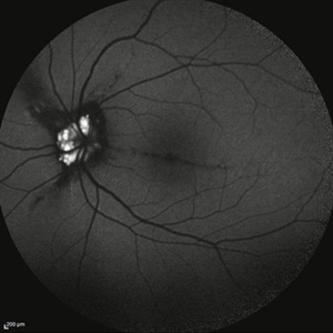
Autofluorescence image of left eye of 53 year female patient with decreased vision in left eye. Patient gives history of multiple joint swellings with multiple dental procedures due to calcification of the roots. She showed type 2 MNV in left eye on OCT and OCTA. Her blood reports showed elevated serum phosphorus (6.4 mg/dl) with normal serum calcium, vitamin D and parathyroid hormone. Her fibroblast growth factor 23 was markedly elevated(>1500RU/ml).
Photographer: Mr Veda Vyas
Condition/keywords: Optic disc drusen and Angiod streaks
-
 Optin Nerve Head Drusen With Angiod Streaks in Hyperphosphatemic Familial Tumoral Calcinosis
Aug 8 2024 by Hemanth Murthy, MBBS, MD, FASRS
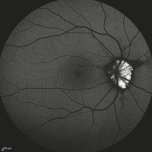
Autofluorescence image of right eye of 53 year female patient with decreased vision in left eye. Patient gives history of multiple joint swellings with multiple dental procedures due to calcification of the roots. She had type2 MNV in left eye demonstrated on OCT and OCTA. Her blood reports showed elevated serum phosphorus (6.4 mg/dl) with normal serum calcium, vitamin D and parathyroid hormone. Her fibroblast growth factor 23 was markedly elevated(>1500RU/ml).
Photographer: Mr Veda Vyas
Condition/keywords: Optic disc drusen and Angiod streaks
-
 Sclerochoroidal Calcification
Jul 5 2024 by Zach Seim
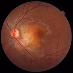
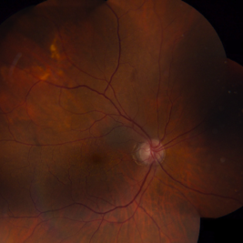
Optos fundus photo of an 81 year old female with Sclerochoroidal Calcification. Patient's VA at presentation was DCC 20/70-1.
Photographer: Zach Seim
Imaging device: Optos California
Condition/keywords: optos, OPTOS CALIFORNIA, sclerochoroidal calcification
-
 Retinoblastoma
Nov 1 2023 by ANKIT JAIN
USG B SCAN image showing hyperechogenic mass lesion with moderate spikes with restricted after movements on dynamic scan. In between high spikes noted suggestive of calcification in a case of Retinoblastoma
Photographer: DR ANKIT JAIN
Condition/keywords: B scan ultrasound, retinoblastoma, ultrasound
-
 Choroidal Osteoma
Jan 3 2022 by Thirumalesh Mochi Basavaraj, MD
Fundus photograph of a young female in her second decade with a choroidal mass lesion with calcification suggestive of choroidal osteomalacia.
Photographer: Putta Swamy, Narayana Nethralaya
Imaging device: Topcon DRI Triton
Condition/keywords: macular choroidal osteoma
-
 Bilateral Calcific Retina Arteriolar Occlusions in a Patient with Metastatic Ovarian Carcinoma
Dec 10 2020 by McGill University Health Centre
47-year-old female with cough and fever. Imaging showed a right pulmonary infiltrate. Transbronchial needle biopsy revealed lymphangitic spread of papillary adenocarcinoma with psammoma bodies. MRI of thyroid, CT of abdomen and pelvis were negative. gynecologic evaluation negative at that time . The patient had bilateral floaters, VA: 20/40 OD and 20/20 OS. Fundus examination showed retinal arteriolar sheathing and a flat choroidal lesion OS and vitritis OD. Fluorescein angiogram showed staining of left superior temporal retinal arterioles and bilateral midperipheral patchy hyperfluorescence at RPE The patient vision in the OD deteriorated to 20/400, and in the OS 20/50. Four months later a new choroidal lesion was diagnosed OS. An abdominal mass consistent with a cystadenoma of the ovary was diagnosed. After a year patient developed systemic metastasis. Autopsy: Metastatic adenocarcinoma to the lung, both adrenals, para-aortic lymph nodes, left hip, right breast, occipital skin, serosal surface of liver, pituitary. In almost all metastatic lesions psammoma bodies were found. Presumptive diagnosis is a primary tumor of the ovary.
Condition/keywords: bilateral, calcification, metastatic adenocarcinoma, retinal arteriolar occlusion
-
 Bilateral Calcific Retina Arteriolar Occlusions in a Patient with Metastatic Ovarian Carcinoma
Dec 10 2020 by McGill University Health Centre
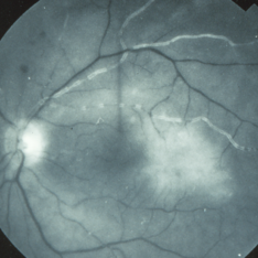
47-year-old female with cough and fever. Imaging showed a right pulmonary infiltrate. Transbronchial needle biopsy revealed lymphangitic spread of papillary adenocarcinoma with psammoma bodies (MRI of thyroid, CT of abdomen and pelvis were negative) gynecologic evaluation negative at that time . The patient had bilateral floaters, VA: 20/40 OD and 20/20 OS. Fundus examination showed retinal arteriolar sheathing and a flat choroidal lesion OS and vitritis OD. Fluorescein angiogram showed staining of left superior temporal retinal arterioles and bilateral midperipheral patchy hyperfluorescence at RPE. The patient vision in the OD deteriorated to 20/400, and in the OS 20/50. Four months later a new choroidal lesion was diagnosed OS. An abdominal mass consistent with a cystadenoma of the ovary was diagnosed. After a year patient developed systemic metastasis. Autopsy: Metastatic adenocarcinoma to the lung, both adrenals, para-aortic lymph nodes, left hip, right breast, occipital skin, serosal surface of liver, pituitary. In almost all metastatic lesions psammoma bodies were found. Presumptive diagnosis is a primary tumor of the ovary.
Imaging device: Fluoroscein angiogram
Condition/keywords: bilateral, calcification, metastatic adenocarcinoma, retinal arteriolar occlusion
-
 Bilateral Calcific Retina Arteriolar Occlusions in a Patient with Metastatic Ovarian Carcinoma
Dec 10 2020 by McGill University Health Centre
47-year-old female with cough and fever. Imaging showed a right pulmonary infiltrate. Transbronchial needle biopsy revealed lymphangitic spread of papillary adenocarcinoma with psammoma bodies (MRI of thyroid, CT of abdomen and pelvis were negative) gynecologic evaluation negative at that time . The patient had bilateral floaters, VA: 20/40 OD and 20/20 OS. Fundus examination showed retinal arteriolar sheathing and a flat choroidal lesion OS and vitritis OD. Fluorescein angiogram showed staining of left superior temporal retinal arterioles and bilateral midperipheral patchy hyperfluorescence at RPE The patient vision in the OD deteriorated to 20/400, and in the OS 20/50. Four months later a new choroidal lesion was diagnosed OS. An abdominal mass consistent with a cystadenoma of the ovary was diagnosed. After a year patient developed systemic metastasis. Autopsy: Metastatic adenocarcinoma to the lung, both adrenals, para-aortic lymph nodes, left hip, right breast, occipital skin, serosal surface of liver, pituitary. In almost all metastatic lesions psammoma bodies were found. Presumptive diagnosis is a primary tumor of the ovary. Histopathologic examination of both eyes disclosed : Bilateral metastatic adenocarcinoma to the vitreous with partially calcified proliferation along internal limiting membrane, OS. Metastatic adenocarcinoma to choroid, OS. Bilateral optic atrophy secondary to retinal arteriolar occlusion with calcification.
Condition/keywords: bilateral, calcification, histopathology, metastatic adenocarcinoma, pathology, retinal arteriolar occlusion
-
 Bilateral Calcific Retina Arteriolar Occlusions in a Patient with Metastatic Ovarian Carcinoma
Dec 10 2020 by McGill University Health Centre
47-year-old female with cough and fever. Imaging showed a right pulmonary infiltrate. Transbronchial needle biopsy revealed lymphangitic spread of papillary adenocarcinoma with psammoma bodies (MRI of thyroid, CT of abdomen and pelvis were negative) gynecologic evaluation negative at that time Patient had bilateral floaters, VA: 20/40 OD and 20/20 OS. Fundus examination showed retinal arteriolar sheathing and a flat choroidal lesion OS and vitritis OD. Fluorescein angiogram showed staining of left superior temporal retinal arterioles and bilateral midperipheral patchy hyperfluorescence at RPE The patient vision in the OD deteriorated to 20/400, and in the OS 20/50. Four months later a new choroidal lesion was diagnosed OS. An abdominal mass consistent with a cystadenoma of the ovary was diagnosed. After a year patient developed systemic metastasis. Autopsy: Metastatic adenocarcinoma to the lung, both adrenals, para-aortic lymph nodes, left hip, right breast, occipital skin, serosal surface of liver, pituitary. In almost all metastatic lesions psammoma bodies were found. Presumptive diagnosis is a primary tumor of the ovary. Histopathologic examination of both eyes disclosed : Bilateral metastatic adenocarcinoma to the vitreous with partially calcified proliferation along internal limiting membrane, OS. Metastatic adenocarcinoma to choroid, OS. Bilateral optic atrophy secondary to retinal arteriolar occlusion with calcification.
Condition/keywords: bilateral, calcification, histopathology, metastatic adenocarcinoma, pathology, retinal arteriolar occlusion
-
 Bilateral Calcific Retina Arteriolar Occlusions in a Patient with Metastatic Ovarian Carcinoma
Dec 10 2020 by McGill University Health Centre
47-year-old female with cough and fever. Imaging showed a right pulmonary infiltrate. Transbronchial needle biopsy revealed lymphangitic spread of papillary adenocarcinoma with psammoma bodies (MRI of thyroid, CT of abdomen and pelvis were negative) gynecologic evaluation negative at that time . The patient had bilateral floaters, VA: 20/40 OD and 20/20 OS. Fundus examination showed retinal arteriolar sheathing and a flat choroidal lesion OS and vitritis OD. Fluorescein angiogram showed staining of left superior temporal retinal arterioles and bilateral midperipheral patchy hyperfluorescence at RPE. The patient vision in the OD deteriorated to 20/400, and in the OS 20/50. Four months later a new choroidal lesion was diagnosed OS. An abdominal mass consistent with a cystadenoma of the ovary was diagnosed. After a year patient developed systemic metastasis. Autopsy: Metastatic adenocarcinoma to the lung, both adrenals, para-aortic lymph nodes, left hip, right breast, occipital skin, serosal surface of liver, pituitary. In almost all metastatic lesions psammoma bodies were found. Presumptive diagnosis is a primary tumor of the ovary. Histopathologic examination of both eyes disclosed : Bilateral metastatic adenocarcinoma to the vitreous with partially calcified proliferation along internal limiting membrane, OS. Metastatic adenocarcinoma to choroid, OS. Bilateral optic atrophy secondary to retinal arteriolar occlusion with calcification.
Condition/keywords: bilateral, calcification, histopathology, metastatic adenocarcinoma, pathology, retinal arteriolar occlusion
-
 Bilateral Calcific Retina Arteriolar Occlusions in a Patient with Metastatic Ovarian Carcinoma
Dec 10 2020 by McGill University Health Centre
47-year-old female with cough and fever. Imaging showed a right pulmonary infiltrate. Transbronchial needle biopsy revealed lymphangitic spread of papillary adenocarcinoma with psammoma bodies (MRI of thyroid, CT of abdomen and pelvis were negative) gynecologic evaluation negative at that time . The patient had bilateral floaters, VA: 20/40 OD and 20/20 OS. Fundus examination showed retinal arteriolar sheathing and a flat choroidal lesion OS and vitritis OD. Fluorescein angiogram showed staining of left superior temporal retinal arterioles and bilateral midperipheral patchy hyperfluorescence at RPE The patient vision in the OD deteriorated to 20/400, and in the OS 20/50. Four months later a new choroidal lesion was diagnosed OS. An abdominal mass consistent with a cystadenoma of the ovary was diagnosed. After a year patient developed systemic metastasis. Autopsy: Metastatic adenocarcinoma to the lung, both adrenals, para-aortic lymph nodes, left hip, right breast, occipital skin, serosal surface of liver, pituitary. In almost all metastatic lesions psammoma bodies were found. Presumptive diagnosis is a primary tumor of the ovary. Histopathologic examination of both eyes disclosed : Bilateral metastatic adenocarcinoma to the vitreous with partially calcified proliferation along internal limiting membrane, OS. Metastatic adenocarcinoma to choroid, OS. Bilateral optic atrophy secondary to retinal arteriolar occlusion with calcification.
Condition/keywords: bilateral, calcification, histopathology, metastatic adenocarcinoma, pathology, retinal arteriolar occlusion
-
 Bilateral Calcific Retina Arteriolar Occlusions in a Patient with Metastatic Ovarian Carcinoma
Dec 10 2020 by McGill University Health Centre
47-year-old female with cough and fever. Imaging showed a right pulmonary infiltrate. Transbronchial needle biopsy revealed lymphangitic spread of papillary adenocarcinoma with psammoma bodies (MRI of thyroid, CT of abdomen and pelvis were negative) gynecologic evaluation negative at that time . The patient had bilateral floaters, VA: 20/40 OD and 20/20 OS. Fundus examination showed retinal arteriolar sheathing and a flat choroidal lesion OS and vitritis OD. Fluorescein angiogram showed staining of left superior temporal retinal arterioles and bilateral midperipheral patchy hyperfluorescence at RPE. The patient vision in the OD deteriorated to 20/400, and in the OS 20/50. Four months later a new choroidal lesion was diagnosed OS. An abdominal mass consistent with a cystadenoma of the ovary was diagnosed. After a year patient developed systemic metastasis. Autopsy: Metastatic adenocarcinoma to the lung, both adrenals, para-aortic lymph nodes, left hip, right breast, occipital skin, serosal surface of liver, pituitary. In almost all metastatic lesions psammoma bodies were found. Presumptive diagnosis is a primary tumor of the ovary. Histopathologic examination of both eyes disclosed : Bilateral metastatic adenocarcinoma to the vitreous with partially calcified proliferation along internal limiting membrane, OS. Metastatic adenocarcinoma to choroid, OS. Bilateral optic atrophy secondary to retinal arteriolar occlusion with calcification.
Condition/keywords: bilateral, calcification, histopathology, metastatic adenocarcinoma, pathology, retinal arteriolar occlusion
-
 Infiltration of the Optic Nerve
May 18 2020 by McGill University Health Centre
The main morphological prognostic factor for retinoblastoma is infiltration of the optic nerve. The invasion of the optic nerve has to be assessed by a pathologist in all cases. The superior calotte is removed in this enucleation specimen to show an extensively exophytic necrotic tumor occupying the vitreous chamber with severe infiltration of the optic nerve (arrowhead). Intratumoral calcification is a hallmark of this tumor and can be seen in the chalky white areas (arrow).
Condition/keywords: infiltration of the optic nerve
-
 Retinoblastoma With Calcifications
Dec 16 2019 by Sophia El Hamichi, MD
A 4-year-old male patient with germline retinoblastoma, treated with intraarterial chemotherapy and thermal transpupillary laser.
Photographer: Abby Orcutt-Hayes, Murray Ocular Oncology and Retina
Condition/keywords: calcification, chemoreduction, retinoblastoma
-
 Retinoblastoma With Calcifications: Fluorescein Angiogram
Dec 16 2019 by Sophia El Hamichi, MD
A 4-yeasr-old male patient with germline retinoblastoma, treated with intraarterial chemotherapy and thermal transpupillary laser.
Photographer: Abby Orcutt-Hayes, Murray Ocular Oncology and Retina
Condition/keywords: fluorescein angiogram (FA), retinoblastoma
-
 Retinal Astrocytic Hamartomas
Dec 3 2019 by Timothy S Fuller, MD
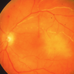
Fundus photograph of an 11-year-old boy with known tuberous sclerosis complex. Note calcification present in large lesion nasal to optic nerve.
Photographer: Tony Abate
Imaging device: Optos
Condition/keywords: astrocytic hamartoma, tuberous sclerosis
-
 Idiopathic Sclerochoroidal Calcification
Aug 12 2019 by Jonathan C. Tsui, MD
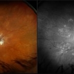
A 73-year-old Caucasian male presents with asymptomatic unilateral idiopathic sclerochoroidal calcification at the right superotemporal arcade. B-scan demonstrated hyperreflectivity of the lesion. Recent electrolyte testing was unremarkable.
Condition/keywords: idiopathic sclerochoroidal calcification
-
 IOL Opacification
Jul 30 2019 by Haider Ali
70-year-old non diabetic, non hypertensive female who had cataract surgery 3 years ago presents in opd with gradually decreasing vision in her left eye. V/A is counting fingers and 1 meters. B scan examination is normal.
Photographer: Dr Haider Ali Chaudhry, Madinah Teaching Hospital
Condition/keywords: IOL calcification, IOL opacification
-
 Calcifications in Retinoblastoma
Apr 2 2019 by Gary R. Cook, MD, FACS
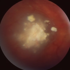
A calcified, necrotic retinoblastoma lesion OD in a 13-month old white male infant with bilateral retinoblastoma.
Condition/keywords: retinoblastoma
-
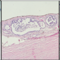
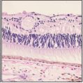
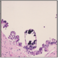
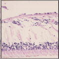
 Retinoblastoma Pathology
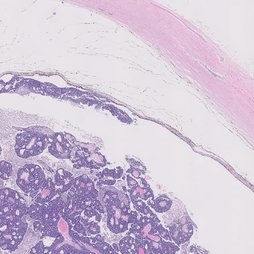
Jan 10 2019 by Rahul Komati, MD
Pathology showing poorly differentiated tumor without classic rosettes; many apoptoses, cystic spaces, calcifications.
Condition/keywords: retinoblastoma
 Loading…