Search results (53 results)
-
 Retinoblastoma Ultrasound
Oct 2 2015 by Aparna Ramasubramanian
Ultrasonography of a retinoblastoma tumor shows hyperreflective echoes suggestive of calcification. It is seen in 90% of retinoblastoma patients and is an important diagnostic sign.
Photographer: Aparna Ramasubramanian
Condition/keywords: A-scan ultrasound, B scan ultrasound, calcification, retinoblastoma
-
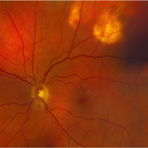
 Retinoblastoma
Oct 5 2012 by Ronald C. Gentile, MD
B scan ultrasonography of the large endophytic retinoblastoma revealing internal hyper-reflective spots consistent with internal calcification.
Photographer: The New York Eye & Ear Infirmary Department of Medical Imaging
Condition/keywords: B scan ultrasound, retinoblastoma
-
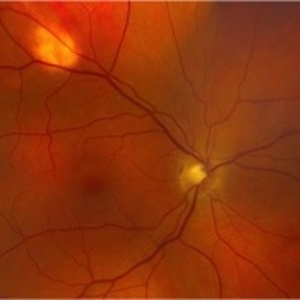
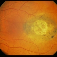
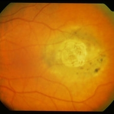
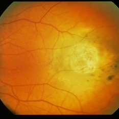
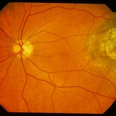
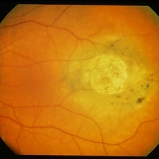
 Choroidal Osteoma and Secondary Choroidal Neovascular Membrane
Sep 21 2012 by Allen Chiang, MD, FASRS
Fundus photograph of a 44-year old woman with a choroidal osteoma complicated by secondary choroidal neovascular membrane, regressed after serial intravitreal bevacizumab injections. The tumor exhibits areas of decalcification.
Imaging device: Topcon
Condition/keywords: choroidal neovascularization (CNV), choroidal osteoma, macular choroidal osteoma
-
---thumb.jpg/image-square;max$300,300.ImageHandler) Floaters
Floaters
Oct 9 2013 by Maurice F. Rabb
KR is a 25 year old white female who presented with a one month history of floaters OD. Past ocular and systemic history were unremarkable. On clinical examination, the visual acuity was 20/20 OU, and the anterior segments were normal. There was a very mild degree of vitreous cell OD, though no cystoid macular edema nor vasculitis. A lobulated white mass was noted overlying the vitreous base inferotemporally OD (thickness 3.3mm). There was no calcification, though prominent cysts were noted on the surface of the lesion. A fluorescein angiogram, echogram, and CT scan were obtained, along with a thorough systemic evaluation.
Condition/keywords: floaters
-
 Sclerochoroidal Calcification
Aug 24 2012 by John S. King, MD
Idiopathic Sclerochoroidal Calcification
Photographer: Kristin Konecki, OcuSight Eye Care Center, Rochester, NY
Condition/keywords: idiopathic sclerochoroidal calcification
-
 Sclerochoroidal Calcification OS
Nov 9 2016 by Courtney Crawford, MD, FACS
55-year-old male with stable choroidal lesions in the superotemporal quadrant of both eyes
Condition/keywords: idiopathic sclerochoroidal calcification
-
---thumb.jpg/image-square;max$300,300.ImageHandler) Floaters
Floaters
Oct 9 2013 by Maurice F. Rabb
KR is a 25 year old white female who presented with a one month history of floaters OD. Past ocular and systemic history were unremarkable. On clinical examination, the visual acuity was 20/20 OU, and the anterior segments were normal. There was a very mild degree of vitreous cell OD, though no cystoid macular edema nor vasculitis. A lobulated white mass was noted overlying the vitreous base inferotemporally OD (thickness 3.3mm). There was no calcification, though prominent cysts were noted on the surface of the lesion. A fluorescein angiogram, echogram, and CT scan were obtained, along with a thorough systemic evaluation.
Condition/keywords: floaters
-
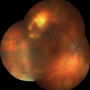
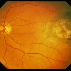
 Retinoblastom Group E
Apr 17 2014 by Susanna S. Park, MD, PhD
Retcam Fundus photograph of a 3-year-old girl with no family history of retinoblastoma noted with a large retinal tumor with calcification filling 70% of globe with diffuse vitreous and subretinal seeding and exudative retinal detachment--unilateral Group E.
Photographer: Ellen Redenbo, University of California Davis Eye Center
Condition/keywords: retinoblastoma, tumor seeding
-
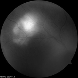
---thumb.jpg/image-square;max$300,300.ImageHandler) Choroidal Osteoma
Choroidal Osteoma
Apr 29 2013 by Subijay Sinha
Ultrasonography shows a curvilinear plaque of high amplitude with shadowing in the juxtapapillary area.
Condition/keywords: calcification, shadow, ultrasound
-
---thumb.jpg/image-square;max$300,300.ImageHandler) Choroidal Osteoma
Choroidal Osteoma
Apr 29 2013 by Subijay Sinha
CT Scan of the sagital section showing the calcification in the eye.
Condition/keywords: calcification, CT scan
-
 Communicating Orbital Cyst
Dec 10 2012 by Yale L. Fisher, MD
Observe orbital cystic structures temporal to optic nerve. Orbital cyst transmits sounds easily. Small optic nerve shadow is visible superior to the cyst in the upper portion of the screen. Strong reflections from choroidal region are consistent with calcification. Tilting of the probe vertically demonstrates communication of the orbital cyst to the vitreous cavity as well as posiible communication to optic nerve.
Condition/keywords: video
-
 Sclerochoroidal Calcification OD
Nov 9 2016 by Courtney Crawford, MD, FACS
55-year-old male with stable choroidal lesions in the superotemporal quadrant of both eyes.
Condition/keywords: idiopathic sclerochoroidal calcification
-
 Sclerochoroidal Calcification AF
Aug 24 2012 by John S. King, MD
AF
Photographer: Kristin Konecki, OcuSight Eye Care Center, Rochester, NY
Condition/keywords: idiopathic sclerochoroidal calcification
-
 Nevus With Calcific Scar
Mar 25 2014 by David Callanan, MD
78-year-old male, nevus with calcific scar.
Condition/keywords: calcification, nevus, scar
-
 Nevus With Calcific Scar
Mar 25 2014 by David Callanan, MD
78-year-old male, nevus with calcific scar.
Condition/keywords: calcification, nevus, scar
-
 Nevus With Calcific Scar
Mar 25 2014 by David Callanan, MD
78-year-old male, nevus with calcific scar.
Condition/keywords: calcification, nevus, scar
-
 Nevus With Calcific Scar
Mar 25 2014 by David Callanan, MD
78-year-old male, nevus with calcific scar.
Condition/keywords: calcification, nevus, scar
-
 Nevus With Calcific Scar
Mar 25 2014 by David Callanan, MD
78-year-old male, nevus with calcific scar.
Condition/keywords: calcification, nevus, scar
-
 Nevus With Calcific Scar
Mar 25 2014 by David Callanan, MD
78-year-old male, nevus with calcific scar.
Condition/keywords: calcification, nevus, scar
-
 Optic Nerve Head Drusen
Dec 10 2012 by Yale L. Fisher, MD
Another movie from Dr. Yale Fisher and the OphthalmicEdge.org Contact B-scan ultrasonography can be of great help in diagnosing buried drusen of the optic nerve head. When drusen are buried deep in the nerve and invisible to white light, ultrasound has proved to be diagnostically helpful. Drusen of the nerve head are excellent reflectors of sound (probably due to the presence of calcification). Drusen themselves have an extensive shadowing effect, but since we are already shooting through the optic nerve shadow, much of this effect is not evident. They stand out like small pebbles on the head of the optic nerve and their characteristic hyper-reflective signal persist at the lowest decibel levels of gain. Also in this eye, scattered vitreous opacities of very low reflectivity are visible, which may be related to advancing age. In younger eyes, the clear vitreous body generally produces no echoes.
Condition/keywords: video
-
 Nevus With Calcific Scar
Mar 25 2014 by David Callanan, MD
78-year-old male, nevus with calcific scar.
Condition/keywords: calcification, nevus, scar
 Loading…