Search results (53 results)
-
 Bilateral Calcific Retina Arteriolar Occlusions in a Patient with Metastatic Ovarian Carcinoma
Dec 10 2020 by McGill University Health Centre
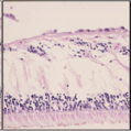
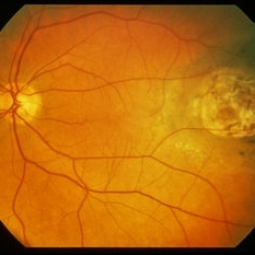
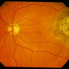
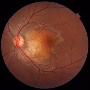
47-year-old female with cough and fever. Imaging showed a right pulmonary infiltrate. Transbronchial needle biopsy revealed lymphangitic spread of papillary adenocarcinoma with psammoma bodies (MRI of thyroid, CT of abdomen and pelvis were negative) gynecologic evaluation negative at that time . The patient had bilateral floaters, VA: 20/40 OD and 20/20 OS. Fundus examination showed retinal arteriolar sheathing and a flat choroidal lesion OS and vitritis OD. Fluorescein angiogram showed staining of left superior temporal retinal arterioles and bilateral midperipheral patchy hyperfluorescence at RPE. The patient vision in the OD deteriorated to 20/400, and in the OS 20/50. Four months later a new choroidal lesion was diagnosed OS. An abdominal mass consistent with a cystadenoma of the ovary was diagnosed. After a year patient developed systemic metastasis. Autopsy: Metastatic adenocarcinoma to the lung, both adrenals, para-aortic lymph nodes, left hip, right breast, occipital skin, serosal surface of liver, pituitary. In almost all metastatic lesions psammoma bodies were found. Presumptive diagnosis is a primary tumor of the ovary. Histopathologic examination of both eyes disclosed : Bilateral metastatic adenocarcinoma to the vitreous with partially calcified proliferation along internal limiting membrane, OS. Metastatic adenocarcinoma to choroid, OS. Bilateral optic atrophy secondary to retinal arteriolar occlusion with calcification.
Condition/keywords: bilateral, calcification, histopathology, metastatic adenocarcinoma, pathology, retinal arteriolar occlusion
-
 Bilateral Calcific Retina Arteriolar Occlusions in a Patient with Metastatic Ovarian Carcinoma
Dec 10 2020 by McGill University Health Centre
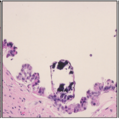
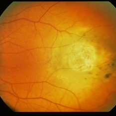
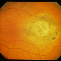
47-year-old female with cough and fever. Imaging showed a right pulmonary infiltrate. Transbronchial needle biopsy revealed lymphangitic spread of papillary adenocarcinoma with psammoma bodies (MRI of thyroid, CT of abdomen and pelvis were negative) gynecologic evaluation negative at that time . The patient had bilateral floaters, VA: 20/40 OD and 20/20 OS. Fundus examination showed retinal arteriolar sheathing and a flat choroidal lesion OS and vitritis OD. Fluorescein angiogram showed staining of left superior temporal retinal arterioles and bilateral midperipheral patchy hyperfluorescence at RPE The patient vision in the OD deteriorated to 20/400, and in the OS 20/50. Four months later a new choroidal lesion was diagnosed OS. An abdominal mass consistent with a cystadenoma of the ovary was diagnosed. After a year patient developed systemic metastasis. Autopsy: Metastatic adenocarcinoma to the lung, both adrenals, para-aortic lymph nodes, left hip, right breast, occipital skin, serosal surface of liver, pituitary. In almost all metastatic lesions psammoma bodies were found. Presumptive diagnosis is a primary tumor of the ovary. Histopathologic examination of both eyes disclosed : Bilateral metastatic adenocarcinoma to the vitreous with partially calcified proliferation along internal limiting membrane, OS. Metastatic adenocarcinoma to choroid, OS. Bilateral optic atrophy secondary to retinal arteriolar occlusion with calcification.
Condition/keywords: bilateral, calcification, histopathology, metastatic adenocarcinoma, pathology, retinal arteriolar occlusion
-
 Bilateral Calcific Retina Arteriolar Occlusions in a Patient with Metastatic Ovarian Carcinoma
Dec 10 2020 by McGill University Health Centre
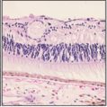
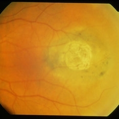
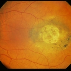
47-year-old female with cough and fever. Imaging showed a right pulmonary infiltrate. Transbronchial needle biopsy revealed lymphangitic spread of papillary adenocarcinoma with psammoma bodies (MRI of thyroid, CT of abdomen and pelvis were negative) gynecologic evaluation negative at that time . The patient had bilateral floaters, VA: 20/40 OD and 20/20 OS. Fundus examination showed retinal arteriolar sheathing and a flat choroidal lesion OS and vitritis OD. Fluorescein angiogram showed staining of left superior temporal retinal arterioles and bilateral midperipheral patchy hyperfluorescence at RPE. The patient vision in the OD deteriorated to 20/400, and in the OS 20/50. Four months later a new choroidal lesion was diagnosed OS. An abdominal mass consistent with a cystadenoma of the ovary was diagnosed. After a year patient developed systemic metastasis. Autopsy: Metastatic adenocarcinoma to the lung, both adrenals, para-aortic lymph nodes, left hip, right breast, occipital skin, serosal surface of liver, pituitary. In almost all metastatic lesions psammoma bodies were found. Presumptive diagnosis is a primary tumor of the ovary. Histopathologic examination of both eyes disclosed : Bilateral metastatic adenocarcinoma to the vitreous with partially calcified proliferation along internal limiting membrane, OS. Metastatic adenocarcinoma to choroid, OS. Bilateral optic atrophy secondary to retinal arteriolar occlusion with calcification.
Condition/keywords: bilateral, calcification, histopathology, metastatic adenocarcinoma, pathology, retinal arteriolar occlusion
-
 Bilateral Calcific Retina Arteriolar Occlusions in a Patient with Metastatic Ovarian Carcinoma
Dec 10 2020 by McGill University Health Centre
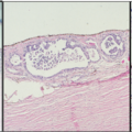
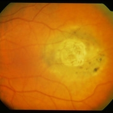
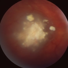
47-year-old female with cough and fever. Imaging showed a right pulmonary infiltrate. Transbronchial needle biopsy revealed lymphangitic spread of papillary adenocarcinoma with psammoma bodies (MRI of thyroid, CT of abdomen and pelvis were negative) gynecologic evaluation negative at that time Patient had bilateral floaters, VA: 20/40 OD and 20/20 OS. Fundus examination showed retinal arteriolar sheathing and a flat choroidal lesion OS and vitritis OD. Fluorescein angiogram showed staining of left superior temporal retinal arterioles and bilateral midperipheral patchy hyperfluorescence at RPE The patient vision in the OD deteriorated to 20/400, and in the OS 20/50. Four months later a new choroidal lesion was diagnosed OS. An abdominal mass consistent with a cystadenoma of the ovary was diagnosed. After a year patient developed systemic metastasis. Autopsy: Metastatic adenocarcinoma to the lung, both adrenals, para-aortic lymph nodes, left hip, right breast, occipital skin, serosal surface of liver, pituitary. In almost all metastatic lesions psammoma bodies were found. Presumptive diagnosis is a primary tumor of the ovary. Histopathologic examination of both eyes disclosed : Bilateral metastatic adenocarcinoma to the vitreous with partially calcified proliferation along internal limiting membrane, OS. Metastatic adenocarcinoma to choroid, OS. Bilateral optic atrophy secondary to retinal arteriolar occlusion with calcification.
Condition/keywords: bilateral, calcification, histopathology, metastatic adenocarcinoma, pathology, retinal arteriolar occlusion
-
 Bilateral Calcific Retina Arteriolar Occlusions in a Patient with Metastatic Ovarian Carcinoma
Dec 10 2020 by McGill University Health Centre
47-year-old female with cough and fever. Imaging showed a right pulmonary infiltrate. Transbronchial needle biopsy revealed lymphangitic spread of papillary adenocarcinoma with psammoma bodies (MRI of thyroid, CT of abdomen and pelvis were negative) gynecologic evaluation negative at that time . The patient had bilateral floaters, VA: 20/40 OD and 20/20 OS. Fundus examination showed retinal arteriolar sheathing and a flat choroidal lesion OS and vitritis OD. Fluorescein angiogram showed staining of left superior temporal retinal arterioles and bilateral midperipheral patchy hyperfluorescence at RPE The patient vision in the OD deteriorated to 20/400, and in the OS 20/50. Four months later a new choroidal lesion was diagnosed OS. An abdominal mass consistent with a cystadenoma of the ovary was diagnosed. After a year patient developed systemic metastasis. Autopsy: Metastatic adenocarcinoma to the lung, both adrenals, para-aortic lymph nodes, left hip, right breast, occipital skin, serosal surface of liver, pituitary. In almost all metastatic lesions psammoma bodies were found. Presumptive diagnosis is a primary tumor of the ovary. Histopathologic examination of both eyes disclosed : Bilateral metastatic adenocarcinoma to the vitreous with partially calcified proliferation along internal limiting membrane, OS. Metastatic adenocarcinoma to choroid, OS. Bilateral optic atrophy secondary to retinal arteriolar occlusion with calcification.
Condition/keywords: bilateral, calcification, histopathology, metastatic adenocarcinoma, pathology, retinal arteriolar occlusion
-
 Bilateral Calcific Retina Arteriolar Occlusions in a Patient with Metastatic Ovarian Carcinoma
Dec 10 2020 by McGill University Health Centre
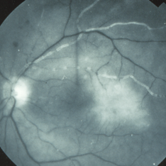
47-year-old female with cough and fever. Imaging showed a right pulmonary infiltrate. Transbronchial needle biopsy revealed lymphangitic spread of papillary adenocarcinoma with psammoma bodies (MRI of thyroid, CT of abdomen and pelvis were negative) gynecologic evaluation negative at that time . The patient had bilateral floaters, VA: 20/40 OD and 20/20 OS. Fundus examination showed retinal arteriolar sheathing and a flat choroidal lesion OS and vitritis OD. Fluorescein angiogram showed staining of left superior temporal retinal arterioles and bilateral midperipheral patchy hyperfluorescence at RPE. The patient vision in the OD deteriorated to 20/400, and in the OS 20/50. Four months later a new choroidal lesion was diagnosed OS. An abdominal mass consistent with a cystadenoma of the ovary was diagnosed. After a year patient developed systemic metastasis. Autopsy: Metastatic adenocarcinoma to the lung, both adrenals, para-aortic lymph nodes, left hip, right breast, occipital skin, serosal surface of liver, pituitary. In almost all metastatic lesions psammoma bodies were found. Presumptive diagnosis is a primary tumor of the ovary.
Imaging device: Fluoroscein angiogram
Condition/keywords: bilateral, calcification, metastatic adenocarcinoma, retinal arteriolar occlusion
-
 Bilateral Calcific Retina Arteriolar Occlusions in a Patient with Metastatic Ovarian Carcinoma
Dec 10 2020 by McGill University Health Centre
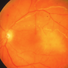
47-year-old female with cough and fever. Imaging showed a right pulmonary infiltrate. Transbronchial needle biopsy revealed lymphangitic spread of papillary adenocarcinoma with psammoma bodies. MRI of thyroid, CT of abdomen and pelvis were negative. gynecologic evaluation negative at that time . The patient had bilateral floaters, VA: 20/40 OD and 20/20 OS. Fundus examination showed retinal arteriolar sheathing and a flat choroidal lesion OS and vitritis OD. Fluorescein angiogram showed staining of left superior temporal retinal arterioles and bilateral midperipheral patchy hyperfluorescence at RPE The patient vision in the OD deteriorated to 20/400, and in the OS 20/50. Four months later a new choroidal lesion was diagnosed OS. An abdominal mass consistent with a cystadenoma of the ovary was diagnosed. After a year patient developed systemic metastasis. Autopsy: Metastatic adenocarcinoma to the lung, both adrenals, para-aortic lymph nodes, left hip, right breast, occipital skin, serosal surface of liver, pituitary. In almost all metastatic lesions psammoma bodies were found. Presumptive diagnosis is a primary tumor of the ovary.
Condition/keywords: bilateral, calcification, metastatic adenocarcinoma, retinal arteriolar occlusion
-
 Calcification of the Retina
Apr 7 2025 by Gustavo Uriel Fonseca Aguirre
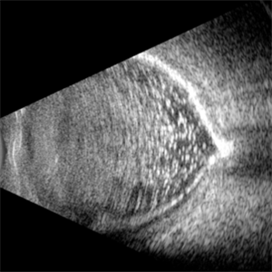
B-mode ultrasound of a vitrectomized eye reveals emulsified silicone oil in the vitreous cavity, retinal detachment, and calcification of the retina and optic nerve head.
Photographer: Gustavo U. Fonseca Aguirre, Hospital Conde de Valenciana, Ciudad de México
Condition/keywords: calcification, Retina detachment, vitrectomy
-
---thumb.jpg/image-square;max$300,300.ImageHandler) Choroidal Osteoma
Choroidal Osteoma
Apr 29 2013 by Subijay Sinha
Ultrasonography shows a curvilinear plaque of high amplitude with shadowing in the juxtapapillary area.
Condition/keywords: calcification, shadow, ultrasound
-
---thumb.jpg/image-square;max$300,300.ImageHandler) Choroidal Osteoma
Choroidal Osteoma
Apr 29 2013 by Subijay Sinha
CT Scan of the sagital section showing the calcification in the eye.
Condition/keywords: calcification, CT scan
-
 Nevus With Calcific Scar
Mar 25 2014 by David Callanan, MD
78-year-old male, nevus with calcific scar.
Condition/keywords: calcification, nevus, scar
-
 Nevus With Calcific Scar
Mar 25 2014 by David Callanan, MD
78-year-old male, nevus with calcific scar.
Condition/keywords: calcification, nevus, scar
-
 Nevus With Calcific Scar
Mar 25 2014 by David Callanan, MD
78-year-old male, nevus with calcific scar.
Condition/keywords: calcification, nevus, scar
-
 Nevus With Calcific Scar
Mar 25 2014 by David Callanan, MD
78-year-old male, nevus with calcific scar.
Condition/keywords: calcification, nevus, scar
-
 Nevus With Calcific Scar
Mar 25 2014 by David Callanan, MD
78-year-old male, nevus with calcific scar.
Condition/keywords: calcification, nevus, scar
-
 Nevus With Calcific Scar
Mar 25 2014 by David Callanan, MD
78-year-old male, nevus with calcific scar.
Condition/keywords: calcification, nevus, scar
-
 Nevus With Calcific Scar
Mar 25 2014 by David Callanan, MD
78-year-old male, nevus with calcific scar.
Condition/keywords: calcification, nevus, scar
-
 Nevus With Calcific Scar
Mar 25 2014 by David Callanan, MD
78-year-old male, nevus with calcific scar.
Condition/keywords: calcification, nevus, scar
-
 Nevus With Calcific Scar
Mar 25 2014 by David Callanan, MD
78-year-old male, nevus with calcific scar.
Condition/keywords: calcification, nevus, scar
-
 Retinoblastoma Ultrasound
Oct 2 2015 by Aparna Ramasubramanian
Ultrasonography of a retinoblastoma tumor shows hyperreflective echoes suggestive of calcification. It is seen in 90% of retinoblastoma patients and is an important diagnostic sign.
Photographer: Aparna Ramasubramanian
Condition/keywords: A-scan ultrasound, B scan ultrasound, calcification, retinoblastoma
-
 Retinoblastoma With Calcifications
Dec 16 2019 by Sophia El Hamichi, MD
A 4-year-old male patient with germline retinoblastoma, treated with intraarterial chemotherapy and thermal transpupillary laser.
Photographer: Abby Orcutt-Hayes, Murray Ocular Oncology and Retina
Condition/keywords: calcification, chemoreduction, retinoblastoma
-
 Calcifications in Retinoblastoma
Apr 2 2019 by Gary R. Cook, MD, FACS
A calcified, necrotic retinoblastoma lesion OD in a 13-month old white male infant with bilateral retinoblastoma.
Condition/keywords: retinoblastoma
-
 Choroidal Osteoma
Jan 3 2022 by Thirumalesh Mochi Basavaraj, MD
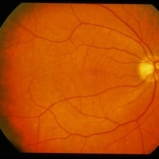
Fundus photograph of a young female in her second decade with a choroidal mass lesion with calcification suggestive of choroidal osteomalacia.
Photographer: Putta Swamy, Narayana Nethralaya
Imaging device: Topcon DRI Triton
Condition/keywords: macular choroidal osteoma
-
 Choroidal Osteoma and Secondary Choroidal Neovascular Membrane
Sep 21 2012 by Allen Chiang, MD, FASRS
Fundus photograph of a 44-year old woman with a choroidal osteoma complicated by secondary choroidal neovascular membrane, regressed after serial intravitreal bevacizumab injections. The tumor exhibits areas of decalcification.
Imaging device: Topcon
Condition/keywords: choroidal neovascularization (CNV), choroidal osteoma, macular choroidal osteoma
-
 Communicating Orbital Cyst
Dec 10 2012 by Yale L. Fisher, MD
Observe orbital cystic structures temporal to optic nerve. Orbital cyst transmits sounds easily. Small optic nerve shadow is visible superior to the cyst in the upper portion of the screen. Strong reflections from choroidal region are consistent with calcification. Tilting of the probe vertically demonstrates communication of the orbital cyst to the vitreous cavity as well as posiible communication to optic nerve.
Condition/keywords: video
 Loading…