 Initializing download.
-
 By McGill University Health Centre
The MUHC-McGill University
Co-author(s): Miguel N. Burnier, Paulina García de Alba Graue, McGill University Health Center-McGill University Ocular Pathology & Translational Research Laboratory - Uploaded on Dec 10, 2020.
- Last modified by Caroline Bozell on Dec 11, 2020.
- Rating
- Appears in
- Bilateral Calcific Retina Arteriolar Occlusions
- Condition/keywords
- metastatic adenocarcinoma, bilateral, retinal arteriolar occlusion, calcification, pathology, histopathology
- Imaging device
- Photo slit lamp biomicroscope
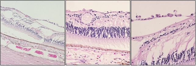
- Description
- 47-year-old female with cough and fever. Imaging showed a right pulmonary infiltrate. Transbronchial needle biopsy revealed lymphangitic spread of papillary adenocarcinoma with psammoma bodies (MRI of thyroid, CT of abdomen and pelvis were negative) gynecologic evaluation negative at that time Patient had bilateral floaters, VA: 20/40 OD and 20/20 OS. Fundus examination showed retinal arteriolar sheathing and a flat choroidal lesion OS and vitritis OD. Fluorescein angiogram showed staining of left superior temporal retinal arterioles and bilateral midperipheral patchy hyperfluorescence at RPE The patient vision in the OD deteriorated to 20/400, and in the OS 20/50. Four months later a new choroidal lesion was diagnosed OS. An abdominal mass consistent with a cystadenoma of the ovary was diagnosed. After a year patient developed systemic metastasis. Autopsy: Metastatic adenocarcinoma to the lung, both adrenals, para-aortic lymph nodes, left hip, right breast, occipital skin, serosal surface of liver, pituitary. In almost all metastatic lesions psammoma bodies were found. Presumptive diagnosis is a primary tumor of the ovary. Histopathologic examination of both eyes disclosed : Bilateral metastatic adenocarcinoma to the vitreous with partially calcified proliferation along internal limiting membrane, OS. Metastatic adenocarcinoma to choroid, OS. Bilateral optic atrophy secondary to retinal arteriolar occlusion with calcification.
---thumb.jpg/image-square;max$79,0.ImageHandler "Toxocariasis Cyst Gross Pathology Specimen")
---thumb.jpg/image-square;max$79,0.ImageHandler "Anterior Hyaloid Fibrovascular Proliferation")


---thumb.jpg/image-square;max$79,0.ImageHandler "Choroidal Osteoma")