Search results (29 results)
-
 Optic Nerve Head Avulsion
Sep 24 2024 by Gustavo Uriel Fonseca Aguirre
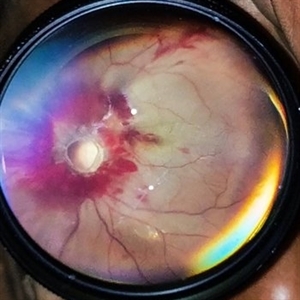
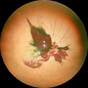
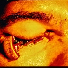
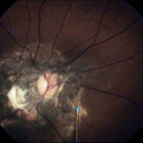
A 14-year-old male with a history of blunt ocular trauma in the right eye presented partial avulsion of the optic nerve head and submacular hemorrhage that was managed with neumatic displacement.
Photographer: Gustavo U. Fonseca Aguirre, Fundación Hospital Nuestra Señora de la Luz, Ciudad de México
Condition/keywords: optic nerve head avulsion
-
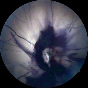
 Optic disc avulsion
Mar 5 2023 by Kalyan Singh
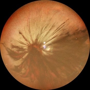
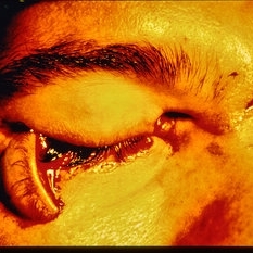
Case of post traumatic optic disc avulsion of young male adult.
Photographer: Kalyan Singh, GSVM medical college, Kanpur
Imaging device: Smartphone (1 plus 10R)
Condition/keywords: optic disc, trauma
-
 Optic nerve Avulsion with Vitreous Hemorrhage
Dec 5 2022 by Vaidehi Sathaye
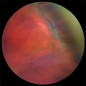
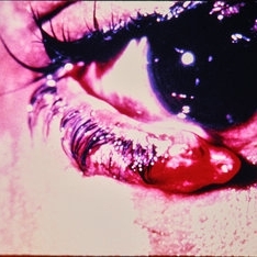
Fundus photograph of LE of a 16 year old male , with optic nerve avulsion with vitreous hemorrhage
Photographer: Dr. Vaidehi Sathaye
Imaging device: Mirante
Condition/keywords: optic nerve avulsion, vitreous hemorrhage
-
 Vitreous Base Avulsion
Feb 21 2022 by Maxwell J Wingelaar, MD
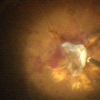
24-year-old male with a vitreous base avulsion with a history of blunt force trauma to the eye
Condition/keywords: vitreous detachment
-
 Vitreous Base Avulsion
Feb 21 2022 by Maxwell J Wingelaar, MD
24-year-old male with a vitreous base avulsion with a history of blunt force trauma to the eye
Condition/keywords: vitreous detachment
-
 Optic Nerve Avulsion with Vitreous Hemorrhage and Pale Retina
Jan 25 2021 by Sham Talati, DOMS
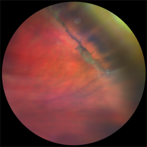
A 30-year-old male presented with history of trauma to RE with NO Perception of light in the affected eye.
Photographer: Dr. Sham Talati,Retina Foundation,Ahmedabad
Imaging device: Nidek Mirante
Condition/keywords: optic nerve, pale retina
-
 Surfboard to OS - Choroidal Rupture, Retinal and Choroidal Avulsion, Retinal Detachment
May 20 2020 by Theodore Leng, MD, MS, FASRS
33-year-old male who sustained a surfboard to the left orbit with orbital fractures, choroidal rupture, nasally and retinal and choroidal avulsion nasally. He also had a horseshoe retinal tear superotemorally and a resultant rhegmatogenous retinal detachment.
Condition/keywords: choroidal avulsion, choroidal rupture, retinal avulsion, retinal detachment with retinal defect
-
 Surfboard to OS - Choroidal Rupture, Retinal and Choroidal Avulsion, Retinal Detachment
May 20 2020 by Theodore Leng, MD, MS, FASRS
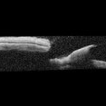
33-year-old male who sustained a surfboard to the left orbit with orbital fractures, choroidal rupture, nasally and retinal and choroidal avulsion nasally. He also had a horseshoe retinal tear superotemorally and a resultant rhegmatogenous retinal detachment. In this OCT image, you can see where the retina broke and nasal to this break, there is fluid under the choroid.
Condition/keywords: choroidal avulsion, choroidal rupture, retinal avulsion
-
 Vitreous Base Avulsion
Sep 19 2019 by Anfisa Ayalon, MD
Fundus picture of a 34-year-old patient with left eye vitreous base avulsion three months after rhegmatogenous retinal detachment repair with circular scleral buckle implantation. Note bucket handle sign and 360 degrees scleral buckle indentation with a flat retina.
Photographer: Anfisa Ayalon, MD., Meir Medical Center, Kfar Saba, Israel.
Imaging device: California, Optos 200 DTX
Condition/keywords: avulsed vitreous base, behind the vitreous base, scleral buckle
-
 Partial Optic Disc Avulsion with Optic Disc Pit
Jul 1 2018 by John S. King, MD
16-year-old with acute loss of vision after blunt finger injury to eye while playing football. Five days post-injury. Vision HM. Decreasing heme and retinal whitening.
Imaging device: Optos
Condition/keywords: traumatic optic neuropathy
-
 Partial Optic Disc Avulsion with Optic Disc Pit
Jul 1 2018 by John S. King, MD
16-year-old with acute loss of vision after blunt finger injury to eye while playing football. He was seen in ED and this is the appearance the next day. Vitreous heme, subhyaloid heme,
Condition/keywords: traumatic optic neuropathy
-
 Partial Optic Disc Avulsion with Optic Disc Pit
Jul 1 2018 by John S. King, MD
16-year-old with acute loss of vision after blunt finger injury to eye while playing football. This photo is three weeks post-injury. Vision HM.
Photographer: Maisee Yang
Imaging device: Topcon
Condition/keywords: epiretinal membrane (ERM), optic disc pit, optic nerve head avulsion, traumatic optic neuropathy
-
 Partial Optic Disc Avulsion with Optic Disc Pit
Jul 1 2018 by John S. King, MD
16-year-old with acute loss of vision after blunt finger injury to eye while playing football. This photo is three weeks post-injury. Vision HM. Retinal striae with subhyaloid heme. Decreased retinal whitening. Peripapillary heme clearing, and temporal optic disc avulsion with optic disc pit can be seen.
Photographer: Maisee Yang
Imaging device: Topcon
Condition/keywords: epiretinal membrane (ERM), optic nerve head avulsion, optic nerve pit, traumatic optic neuropathy
-
 Post Traumatic Optic Nerve Head Avulsion
Nov 18 2017 by Vishal Agrawal, MD, FRCS,FACS,FASRS
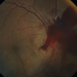
Right eye fundus picture of a 24-year-old male patient who suffered blunt trauma 7 days back with a wooden stick . He presented with NLP vision with a non reacting dilated pupil. Fundus montage picture shows ONH avulsion,CRAO,peripapillary resolving hemorrhages and cicatricial tissue at the edge.
Photographer: Vishal Agrawal, MD, SMS Medical College, Jaipur, India
Imaging device: Zeiss 524
Condition/keywords: avulsion, central retinal artery occlusion (CRAO)
-
 Optic Nerve Head Avulsion
Sep 4 2017 by Shachi Desai
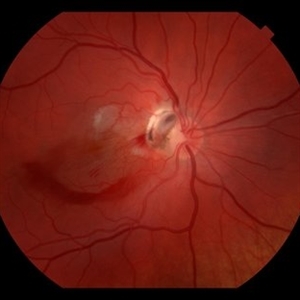
Fundus photograph of 35-year-old male with history of trauma from metal rod during road traffic accident. Patient presented with complaint of complete loss of vision in right eye. Relative afferent pupillary defect and absence of light perception were suggestive of optic nerve involvement. On ophthalmoscopic examination, media was hazy due to vitreous hemorrhage. There was large retinal tear surrounding the probable position of optic nerve head with edematous ischemic retina. Excavated area with hemorrhage at optic nerve head was suggestive of optic nerve head avulsion.
Photographer: Dr Shachi Desai
Imaging device: zeiss visucam
Condition/keywords: blunt trauma, optic nerve head, trauma
-
 Acute Traumatic Optic Nerve Avulsion
Feb 19 2016 by Mahdi Mwas
Fundus photograph of a 24-year-old gentleman, involved in a road traffic accident resulting in left no perception of light.
Photographer: Mahdi Mwas, FRCS, DRCOphth, Jordan
Condition/keywords: optic nerve head avulsion
-
 Traumatic Optic Nerve Avulsion
Jul 16 2015 by Mehul A Shah
A 21-year-old male presented to outdoor with history of blunt trauma and loss of vision on examination we found anterior segment to be normal and posterior segment had this picture.
Photographer: Mehul Shah, Drashti Netralaya
Imaging device: FF450 plus zeiss
Condition/keywords: blunt trauma, optic neuropathy
-
 Traumatic optic nerve avulsion
Apr 23 2015 by Mehul A Shah
30-year-old male presented with blunt ocular trauma following vehicular accident, and lost vision on examination fundus picture is displayed in image.
Photographer: Mehul Shah, Drashti Netralaya
Imaging device: Zeiss FF450plus
Condition/keywords: optic nerve head avulsion, traumatic optic neuropathy
-
 Trauma
-
 Trauma
-
 Trauma
-
 ON Avulsion
Dec 11 2014 by H. Michael Lambert, MD
Optic Nerve Avulsion
Condition/keywords: optic nerve head avulsion
-
 Endstage Diabetic Traction Retinal Detachment
Nov 1 2014 by Maria Stephanie R. Jardeleza, MD
Surgical photograph of optic disc avulsion secondary to extensive diabetic traction retinal membranes over the optic nerve head.
Photographer: Maria Stephanie R. Jardeleza, M.D., UTHSCSA-Texas Diabetes Institute
Imaging device: Zeiss Lumera Operating Microscope
Condition/keywords: proliferative diabetic retinopathy (PDR), tractional retinal detachment
-
 Optic Nerve Head Avulsion
Sep 15 2014 by Mehul A Shah
A 30-year-old male patient met vehicular accident and found to have optic nerve head avulsion with scarring.
Photographer: Drashti Netralaya,Dahod
Imaging device: Zeiss ff450
Condition/keywords: optic nerve head avulsion
-
 Optic Nerve Head Avulsion
Sep 15 2014 by Mehul A Shah
A 30-year-old male presented with loss of vision following blunt trauma.
Photographer: Drashti Netralaya,Dahod
Imaging device: Zeiss ff450
Condition/keywords: optic nerve head avulsion
 Loading…