 Initializing download.
-
 By John S. King, MD
Retina Associates, PA
Co-author(s): Ahmed A. Sallam, M.D., Ph.D., Harvey and Bernice Jones Eye Institute at the University of Arkansas for Medical Sciences (UAMS) - Uploaded on Jul 1, 2018.
- Last modified by Caroline Bozell on Jul 5, 2018.
- Rating
- Appears in
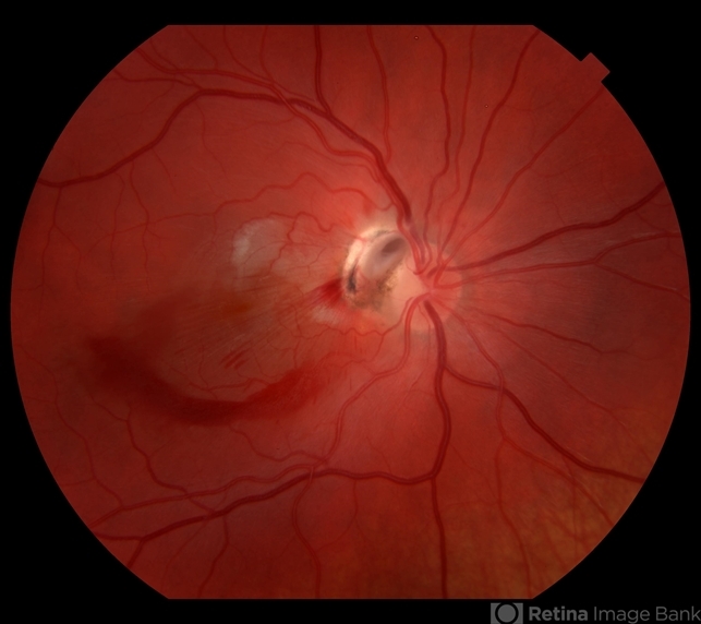
- Optic Disc Avulsion
- Condition/keywords
- optic nerve head avulsion, traumatic optic neuropathy, optic nerve pit, epiretinal membrane (ERM)
- Photographer
- Maisee Yang
- Imaging device
-
Fundus camera
Topcon - Description
- 16-year-old with acute loss of vision after blunt finger injury to eye while playing football. This photo is three weeks post-injury. Vision HM. Retinal striae with subhyaloid heme. Decreased retinal whitening. Peripapillary heme clearing, and temporal optic disc avulsion with optic disc pit can be seen.


---thumb.JPG/image-square;max$79,0.ImageHandler "ERM")
---thumb.jpg/image-square;max$79,0.ImageHandler "ERM")