Search results (2010 results)
-
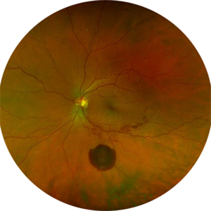
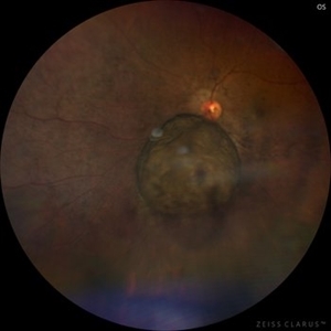
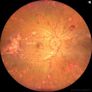 Leukemic Retinopathy and Optic Neuropathy
Aug 25 2025 by Elysse Tom, MD
Fundus photo of a 45-year-old woman with chronic myeloid leukemia.
Condition/keywords: leukemia, leukemic infiltration, Leukemic optic neuropathy
-
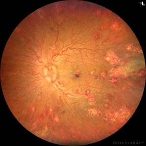 Leukemic Retinopathy and Optic Neuropathy
Aug 25 2025 by Elysse Tom, MD
Fundus photo of a 45-year-old woman with chronic myeloid leukemia.
Condition/keywords: leukemia, leukemic infiltration, Leukemic optic neuropathy
-
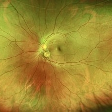
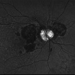 Optic Disc Drusen
Aug 20 2025 by Drew Mitchell
Fundus Autofluorescence photo of an 86 year old woman with neovascular AMD with active CNV and optic disc drusen.
Photographer: Drew Mitchell OCT-C
Imaging device: Optos California
Condition/keywords: fundus autofluorescence (FAF), neovascular age-related macular degeneration (AMD), optic disc drusen, OPTOS
-
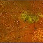 Optic Disc Drusen
Aug 20 2025 by Drew Mitchell
Optos color photo of a 86 year old woman with neovascular AMD with active CNV and optic disc drusen.
Photographer: Drew Mitchell OCT-C
Imaging device: Optos California
Condition/keywords: color photo, optic disc drusen, OPTOS
-
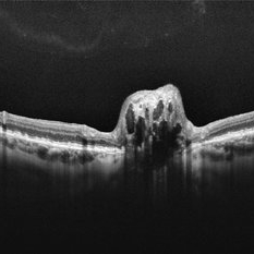 Optic Disc Drusen
Aug 20 2025 by Drew Mitchell
HD 1 line 100x scan through optic disc drusen. ODD are defined as Hyporeflective structures with a full or partial hyperreflective margin.
Photographer: Drew Mitchell OCT-C
Imaging device: Zeiss Cirrus 6000
Condition/keywords: OCT, optic disc drusen
-
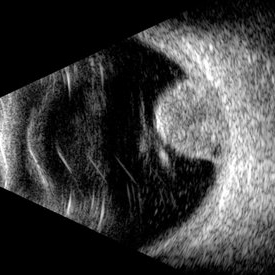
 Posterior Staphyloma + ON-Coloboma
Aug 20 2025 by Gustavo Uriel Fonseca Aguirre
This axial B-scan reveals a highly myopic eye with a posterior staphyloma and an associated optic nerve coloboma. The staphyloma appears as a deep scleral outpouching adjacent to the optic disc, while the coloboma demonstrates a focal posterior excavation with retrobulbar extension.
Photographer: Gustavo U. Fonseca Aguirre, Hospital Conde de Valenciana, Ciudad de México
Condition/keywords: optic nerve coloboma, posterior staphyloma
-
 Goldmann-Favre Syndrome
Aug 19 2025 by Debarun Sharma
Fundus photograph of a 17 year-old female showing circumferential nummular opacities surrounding the vascular arcades. Fundus autoflourescence shows hypo-autoflourescent circumferential opacities with hyper-autoflourescent ring surrounding macula. Left eye also shows hyper-autoflourescent lesion on the optic nerve head suggestive of astrocytic hamartoma. ERG showed reduced cone response with extinguished rod response. OCT showed schisis of macular area. These features are suggestive of Goldmann-Favre Syndrome.
Photographer: Dr. Debarun Sharma, Sri Sankardeva Nethralaya, Guwahati
Imaging device: Optos
Condition/keywords: Goldmann-Favre Syndrome
-
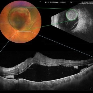 Choroidal Melanoma
Aug 19 2025 by JEFFERSON R SOUSA, Tecg.º (Biomedical Systems Technology)
A 54-year-old woman with progressive visual acuity loss in her left eye was admitted to the institution with a significant elevated lesion in the upper arch with macular involvement, confirmed by wide-angle fundus photography, ultrasound, and optical coherence tomography.
Photographer: JEFFERSON ROCHA DE SOUSA - Retinal Department at Lens Oftalmologia, Sao Paulo-Brazil
Imaging device: Clarus 700 - Zeiss, composite of four 135 degree images.
Condition/keywords: melanoma
-
 Amelanotic Melanoma
Aug 12 2025 by César Adrián Gómez Valdivia, MD
This FAF image reveals a hypoautofluorescent mass with areas of dense hyperautofluorescent stippling—a classic pattern suggestive of an amelanotic choroidal melanoma. Amelanotic melanoma is a rare variant of uveal melanoma, accounting for only a minority of cases. Unlike pigmented melanomas, these lesions lack melanin, making them more challenging to detect on conventional color fundus imaging. FAF Characteristics: • Central hypoautofluorescence: due to loss or compression of the RPE • Peripheral hyperautofluorescent speckling: consistent with lipofuscin accumulation or RPE disruption • Often associated with subretinal fluid or orange pigment seen clinically Location: Juxtapapillary, with potential optic nerve involvement—a factor that complicates both diagnosis and
Photographer: @eyemissu2
Imaging device: California ICG OPTOS
Condition/keywords: amelanotic melanoma
-
 Persistent Hyperplastic Primary Vitreous Associated With Retrolental Fibrovascular Membrane
Aug 8 2025 by Pablo Angel Garcia Uribe
A 12-year-old Mexican male, asymptomatic, referred for evaluation after incidental finding of partial leukocoria on routine ophthalmologic examination. Slit-lamp evaluation revealed a fibrovascular retrolental membrane without evidence of retinal traction, associated with a fibrous stalk connecting to the optic disc. The stalk showed near-complete involution, consistent with a remnant of persistent fetal vasculature (posterior type).
Photographer: Pablo Angel García-Uribe, Clínica Oftalmológica Salauno, Mexico City
Condition/keywords: Persistent Hyperplastic Primary Vitreous Fibrovascular membrane
-
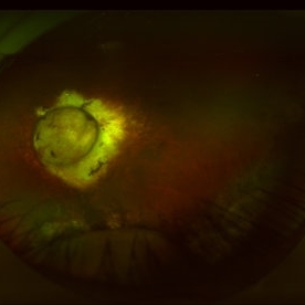
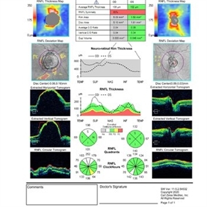 Optic Disc Melanocytoma Hyperpigmented Magnocellular Nevus of the Optic Disk (HMNOD)
Aug 5 2025 by SHRADDHA ASHOK CHANDORKAR, DNB DO FVRS
OCT DISC image of a case of optic nerve melanocytoma.
Photographer: Dr.Shraddha A Chandorkar
Imaging device: zeiss
Condition/keywords: optic disc melanocytoma
-
 Optic Disc Melanocytoma USG Measurements
Aug 5 2025 by SHRADDHA ASHOK CHANDORKAR, DNB DO FVRS
B Scan showing measurements of mass in a case of optic nerve melanocytoma.
Photographer: Dr.Shraddha A Chandorkar
Condition/keywords: b scan
-
 Black Swan - Optic Disc Melanocytoma
Aug 5 2025 by SHRADDHA ASHOK CHANDORKAR, DNB DO FVRS
Just like the Black Swan which signifies an event that comes as a surprise, can have a major effect, and is often inappropriately rationalized after the fact with the benefit of hindsight, a 50 yr old presbyopic lady came to OPD with complains of diminution of vision - BCVA being 6/6 N6 in both eyes. Fundus examination revealed a pigmented nodule covering the optic disc .In most cases, fluorescein angiography of a melanocytoma of the optic disk demonstrates hypofluorescence throughout the angiogram. OCT disc showed elevated lesion, OCT macula normal and USG B scan with measurements were done to corroborate the posterior extension and to note increase in size if any on follow ups, perimetry was done to check for any field defects. All tests seemingly within normal limits - Patient was counselled and asked for 6 monthly follow up. Optic Disc Melanocytoma usually unilateral known to be a benign lesion that carries an excellent prognosis, the malignancy of this specific condition is rare 1-2%. The mean age at diagnosis of optic disk melanocytoma is 50 years with a median of 52 and range of 1–91 years. It is possible that melanocytoma is a congenital lesion but may not become clinically apparent until later in life, perhaps due to acquisition of pigment in a previously amelanotic lesion.
Photographer: Dr.Shraddha A. Chandorkar
Imaging device: topcon
Condition/keywords: optic disc melanocytoma
-
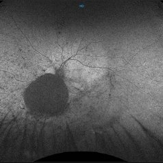
 Large Subhyaloid Hemorrhage
Jul 11 2025 by Jessilla Phou
This is a fundus photograph depicting a large subhyaloid hemorrhage in the mid periphery of the left eye. The patient, a 53-year-old female, presented with a sudden onset of floaters, headache, and blurred vision. The image also demonstrates associated optic disc hemorrhage, vitreous hemorrhage, retinal hemorrhage, and venous tortuosity. Despite the extensive workup performed and the severity of the hemorrhage, no underlying cause was determined.
Photographer: Jessilla Phou
Imaging device: Optos California
Condition/keywords: fundus photograph, optic disc hemorrhage, retinal hemorrhage, venous tortuosity, vitreous hemorrhage
-
 Pseudoduplication of the Optic Disc
Jul 9 2025 by Hrishikesh Naik, MS
A peripapillary colobomatous pseudo-duplication of the optic disc as seen in an asymptomatic 23 year old female with myopia referred for routine retinal periphery screening. Rest retinal exam was normal. Duplication of the optic disc can be classified as either true duplication or pseudoduplication, both of which are rare clinical conditions. Pseudodoubling of the optic disc is commonly caused by optic disc or peripapillary colobomas, characterized by a circumscribed, disc-like lesion with radiating vessels but only one normal optic nerve. A few cases have involved pathological myopia, moderate myopia, proliferative diabetic retinopathy and CHARGE syndrome. The lesion is often found inferior to the normal optic disc. The patient was advised regular follow ups.
Photographer: Hrishikesh Naik
Imaging device: Optos Daytona
Condition/keywords: Coloboma, Pseudoduplication of optic disc
-
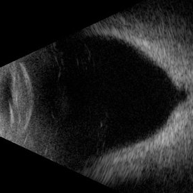
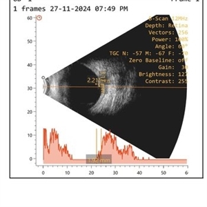
 Choroidal Melanoma (USG)
Jul 5 2025 by Gustavo Uriel Fonseca Aguirre
This B-mode transverse ultrasound scan reveals a mushroom-shaped choroidal tumor in the inferior nasal quadrant adjacent to the optic nerve head. The lesion appears solid with homogeneous internal reflectivity and is associated with minimal surrounding subretinal fluid and scleral excavation. It measures 6.54 mm in height × 7.52 mm in base diameter (transverse view) and extends 9.52 mm longitudinally. The vitreous contains abundant punctate opacities consistent with pigment dispersion. The retina and choroid remain attached elsewhere.
Photographer: Gustavo U. Fonseca Aguirre, Hospital Conde de Valenciana, Ciudad de México
Condition/keywords: choroidal melanoma
-
 Choroidal Hemangioma (AF)
Jul 5 2025 by Gustavo Uriel Fonseca Aguirre
This wide-field fundus autofluorescence image demonstrates a mushroom-shaped choroidal melanoma adjacent to the optic nerve head, exhibiting hypo-autofluorescence (melanin). Vitreous pigment dispersion (tobacco dust sign) is evident, indicating tumor activity.
Photographer: Gustavo U. Fonseca Aguirre, Hospital Conde de Valenciana, Ciudad de México
Condition/keywords: choroidal melanoma
-
 Choroidal Melanoma
Jul 5 2025 by Gustavo Uriel Fonseca Aguirre
This 50° central fundus photograph reveals a mushroom-shaped choroidal melanoma adjacent to the optic nerve head. The lesion demonstrates characteristic pigmentation with overlying vitreous pigment dispersion (tobacco dust sign).
Photographer: Gustavo U. Fonseca Aguirre, Hospital Conde de Valenciana, Ciudad de México
Condition/keywords: choroidal melanoma
-
 Prepapillary Vascular Loop
Jul 4 2025 by KANWALJEET HARJOT MADAN, M.S. (Ophthalmology); FAICO (Vitreous - Retina)
This is the fundus picture of right eye of a young 32 years female depicting pre papillary vascular loop. A prepapillary vascular loop is a congenital anomaly of the optic disc that presents as an elevated and twisted bundle of vessels projecting into the vitreous cavity. It is a benign condition, usually unilateral but can be bilateral. It is asymptomatic and discovered during routine eye examination. This anomaly can sometimes cause complications like branch retinal artery occlusion, vitreous hemorrhage, or sub retinal hemorrhage.
Photographer: Dr. Kanwaljeet Harjot Madan, Thind Eye Hospital, Jalandhar City (Punjab) INDIA.
Imaging device: Zeiss Fundus Camera
Condition/keywords: branch retinal artery occlusion (BRAO), optic disc, Prepapillary Vascular Loop, SUB RETINAL HEMORRHAGE, Vitreous hemorrhage
-
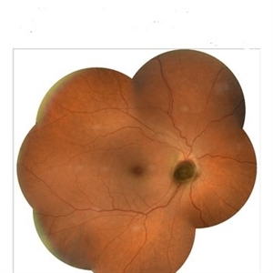
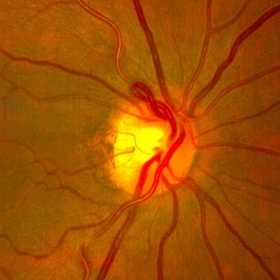
 Coloboma of Optic Disc
Jun 24 2025 by yao zhang
Coloboma of optic disc.
Photographer: Yao Zhang,TongUniversit
Condition/keywords: coloboma of optic disc
-
 Serpiginous Choroidopathy
Jun 23 2025 by César Adrián Gómez Valdivia, MD
Fundus photograph of a 29 year-old female patient diagnosed with Serpiginous Choroidopathy. Finings were bilateral. The most common complication of SC is choroidal neovascularization affecting up to 35% of patients. Other reported complications are subretinal fibrosis, cystoid macular edema, branch vein occlusion, serous retinal detachment, optic disc neovascularization ,and anterior uveitis.
Photographer: @eyemissu2
Imaging device: TOPCON TRC-50DX
Condition/keywords: serpiginous choroiditis
-
 Serpiginous Choroidopathy
Jun 23 2025 by César Adrián Gómez Valdivia, MD
Fundus photograph of a 29 year-old female patient diagnosed with Serpiginous Choroidopathy. Finings were bilateral. The most common complication of SC is choroidal neovascularization affecting up to 35% of patients. Other reported complications are subretinal fibrosis, cystoid macular edema, branch vein occlusion, serous retinal detachment, optic disc neovascularization, and anterior uveitis.
Photographer: @eyemissu2
Imaging device: California ICG OPTOS
Condition/keywords: serpiginous choroiditis
-
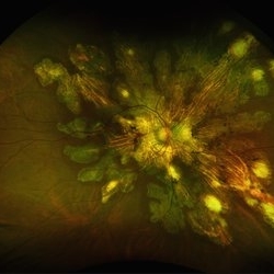
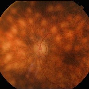 Birdshot Retinochoroidopathy
Jun 18 2025 by César Adrián Gómez Valdivia, MD
Fundus photograph of a 86 YO female patient diagnosed with Birdshot Retinochoroidopathy. Characteristically multifocal cream-colored or yellow-orange, oval or round lesions that emerge from around the optic nerve can be appreciated.
Photographer: @eyemissu2
Imaging device: TOPCON TRC-50DX
Condition/keywords: Birdshot Retinochoroidopathy
-
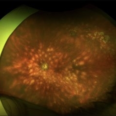 Birdshot Retinochoroidopathy
Jun 18 2025 by César Adrián Gómez Valdivia, MD
Fundus photograph of a 86 YO female patient diagnosed with Birdshot Retinochoroidopathy. Characteristically multifocal cream-colored or yellow-orange, oval or round lesions that emerge from around the optic nerve can be appreciated.
Photographer: @eyemissu2
Imaging device: California ICG OPTOS
Condition/keywords: Birdshot Retinochoroidopathy
-
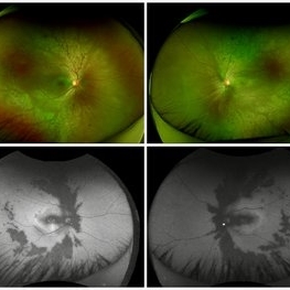
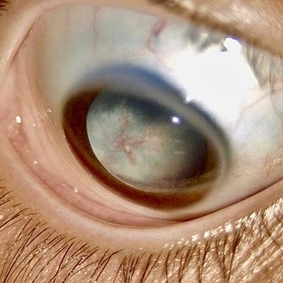
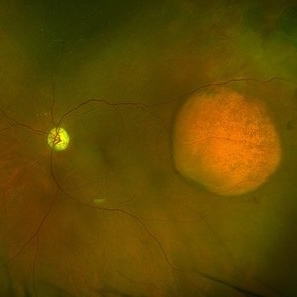 Amelanotic Choroidal Melanoma with Optic Atrophy
Jun 11 2025 by Aditya S Kelkar, MS, FRCS, FASRS,FRCOphth
Fundus photograph of a 64-year-old woman with optic atrophy and amelanotic choroidal melanoma temporal to the macula.
Photographer: Dr Harsh Jain, National Institute of Ophthalmology
Imaging device: Optos Daytona
Condition/keywords: amelanotic melanoma, optic atrophy
 Loading…