Search results (2010 results)
-
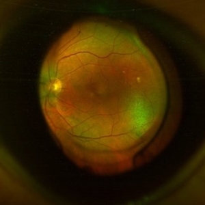
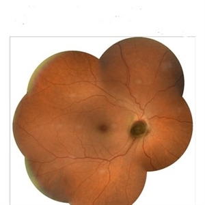
 Optic Nerve Melanocytoma
Mar 14 2024 by César Adrián Gómez Valdivia, MD
Benign neoplasm with seldom malignant transformation.
Photographer: Erika Paulina Ornelas Cazares
Imaging device: TOPCON TRC-50DX
Condition/keywords: disc, Melanocytoma, Nerve, Optic, optic disc melanocytoma
-
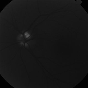
 Optic Nerve Metastasis
May 26 2025 by César Adrián Gómez Valdivia, MD
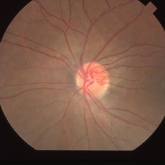
Fundus photograph of a 62 year-old woman with breast cancer history presented to the ER with decreased visual acuity. Optic nerve metastasis were found. Findings were bilateral.
Photographer: @eyemissu2
Imaging device: California ICG OPTOS
Condition/keywords: nerve, Optic, retinal metastasis
-
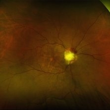
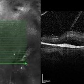
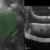
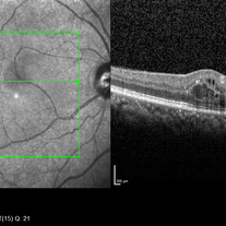
 3D OCT of juxtapapillary melanoma
May 15 2020 by Sophia El Hamichi, MD
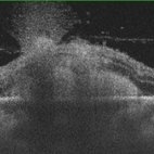
A 63-year-old male with juxtapapillary melanoma of the right eye. Visual acuity at presentation was 20/25 OD. Patient treated with brachytherapy Iodine125 plaque
Photographer: Belinda Rodriguez
Condition/keywords: optical coherence tomography (OCT)
-
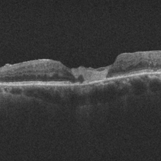
 Acquired Optic Pit Maculopathy
Aug 20 2014 by Andree Henaine-Berra, MD
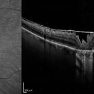
Optical coherence tomography of the left eye of a 60-year-old man with an acquired optic pit maculopathy and glaucoma. The image shows an enlarged optic disc cup and a macular serous detachment.
Photographer: Andree Henaine-Berra. Asociacion Para Evitar la Ceguera en Mexico. Mexico City.
Imaging device: Heidelberg Spectralis
Condition/keywords: glaucoma, maculopathy, optic pit
-
 Acquired Optic Pit Maculopathy
Aug 20 2014 by Andree Henaine-Berra, MD
Optical coherence tomography of the left eye of a 60-year-old man with an acquired optic pit maculopathy and glaucoma. The image shows subretinal fluid extending to the optic nerve and schisis of the outer retinal layers.
Photographer: Andree Henaine-Berra. Asociacion Para Evitar la Ceguera en Mexico. Mexico City.
Imaging device: Heidelberg Spectralis
Condition/keywords: glaucoma, maculopathy, optic pit
-
 Acute Traumatic Optic Nerve Avulsion
Feb 19 2016 by Mahdi Mwas
Fundus photograph of a 24-year-old gentleman, involved in a road traffic accident resulting in left no perception of light.
Photographer: Mahdi Mwas, FRCS, DRCOphth, Jordan
Condition/keywords: optic nerve head avulsion
-
 Advanced PDR
Sep 1 2014 by Hamid Ahmadieh, MD
OCT image of the right eye of a 50-year-old woman with advanced PDR.
Photographer: Soodabeh Fooladian, Negah Eye Center, Tehran, Iran
Condition/keywords: optical coherence tomography (OCT), proliferative diabetic retinopathy (PDR)
-
 Advanced PDR
Sep 1 2014 by Hamid Ahmadieh, MD
OCT image of the left eye of a 50-year-old woman with advanced PDR.
Photographer: Soodabeh Fooladian, Negah Eye Center, Tehran, Iran
Condition/keywords: optical coherence tomography (OCT), proliferative diabetic retinopathy (PDR)
-
 Amniotic-Membrane Grafted Macular Hole
Oct 25 2023 by Jessica Hampton, BS
Optical-coherence tomography image of a 67-year old woman with a recurrent, chronic full-thickness macular hole in the left eye repaired with an amniotic membrane graft, seen at 2 years follow up.
Photographer: Dr. Diana Do, Stanford Medicine, Byers Eye Institute
Condition/keywords: amniotic membrane graft, full thickness macular hole
-
 Aneurysm OCT
Sep 17 2019 by Zachary M Bodnar, MD
Aneurysm OCT
Condition/keywords: optical coherence tomography (OCT), retinal arterial macroaneurysm
-
 Angioid streak-associated choroidal neovasclar membranes
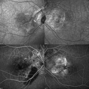
Dec 27 2016 by Young Hee Yoon, MD, PhD
Optical coherence tomogaphs of an 67-year-old woman with CNVM associated with angioid streak in both eyes. (upper row : IR image) Irregular crak-like streaks (lower row : FAG image) Block fluorescence due to subretinal hemorrhage in her right eye and classic CNV in her left eye.
Photographer: Young Hee Yoon, University of Ulsan, Asan Medical Center, Seoul, Korea
Imaging device: Spectralis
Condition/keywords: angioid streaks, choroidal neovascularization (CNV)
-
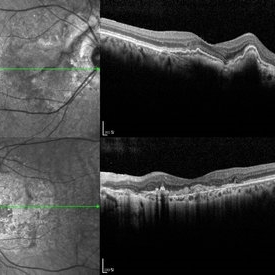 Angioid Streak-Associated Choroidal Neovasclar Membranes
Dec 27 2016 by Young Hee Yoon, MD, PhD
Optical coherence tomogaphs of an 74-year-old woman who received several anti-VEGF injections due to CNV associated with angioid streak in both eyes. There are diffuse CNVM in her right eye and subretinal scar in her left eye. Note the irregular crack in IR image of right eye and the focal Bruch's membrane dehiscence in corresponding B-scan image.
Photographer: Young Hee Yoon, University of Ulsan, Asan Medical Center, Seoul, Korea
Imaging device: Spectralis
Condition/keywords: angioid streaks, choroidal neovascularization (CNV)
-
 AngioOCT Normal Widefield Scan
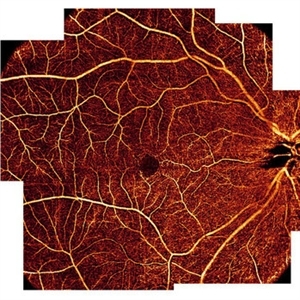
May 8 2015 by Timur Shaimov
Optical coherence tomography angiography of a 28-year-old woman without any macular pathology. Seven 6x6mm angioOCT EnFace images used to merge into widefield view. We used the Optovue RTVue XR Avanti (Optovue, USA) optical coherence tomography system with split-spectrum amplitude decorrelation angiography algorithm (SSADA).
Photographer: Timur Shaimov
Imaging device: Optovue RTVue XR Avanti
Condition/keywords: optical coherence tomography (OCT)
-
 Antero-Posterior Glance
Nov 5 2023 by rahul saradge
image through the principle axis with visibility of all structure in pathway .
Photographer: Optom Rahul , Isha Netralaya
Condition/keywords: optical, Optos, posterior chamber intraocular lens (PCIOL)
-
 Astrocytic Hamartoma
Apr 30 2015 by Mariam A Al-Feky, MD
A 15-year-old boy with history of seizures controlled on treatment. C/O: OD painless DV 10/7 ago (accidental discovery) O/E: BCVA OD: 6/60 ,, OS 6/6. AS: NAD OU. Pupil: RRR no RAPD OU. Fundus examination OD showed a retinitis like lesion with an overlying corkscrew vessel well evident on FFA with late leakage and CSR and OCT through the retinitis like lesion shows diffuse hypereflective thickeninig in the superficial NFL. Thorough history taking revealed that patient has seizures and MRI lesions suggestive of tuberous sclerosis. So this is exudative hamartoma secondary to tuberous sclerosis with marked resolution after single IVI of Lucentis. Retinitis like lesion with corkscrew vessels in FFA is typical together with the homogenous hypereflective thickening in the NFL.
Photographer: Mariam AL-Feky
Imaging device: Optical coherence tomography
Condition/keywords: astrocytic hamartoma
-
 Autofluorescence of Optic Disc Drusen
Mar 2 2014 by Homayoun Tabandeh, MD, FASRS
Autofluorescence of optic disc drusen.
Condition/keywords: optic disc drusen
-
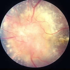 Bilateral Optic Nerve Involvement in Sarcoidosis
Feb 25 2013 by Henry J. Kaplan, MD
Optic nerve head granuloma of sarcoidosis with severe infiltration and exudation in the left eye of the same patient #2.
Condition/keywords: bilateral involvement, sarcoid granuloma
-
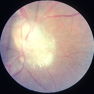 Bilateral Optic Nerve Involvement in Sarcoidosis
Feb 25 2013 by Henry J. Kaplan, MD
Optic nerve granuloma of sarcoidosis in the right eye of a patient with bilateral involvement #1. Left eye is in the following slide.
Condition/keywords: bilateral involvement, sarcoid granuloma
-
 Bilateral Optic Nerve Pits - ON OCTs
Sep 18 2012 by Pauline T Merrill, MD, FASRS
Optic nerve OCTs of a 77-year-old woman with bilateral optic nerve pits and glaucoma, stable over 20 years.
Photographer: Karen Parque, Illinois Retina Associates, Chicago, IL
Imaging device: Zeiss Cirrus
Condition/keywords: optic nerve pit
-
 Binder4 P25 Slide140
Feb 20 2013 by From the Collections of Thomas M. Aaberg, MD and Thomas M. Aaberg Jr., MD
40-year-old white female.
Condition/keywords: optic nerve pit
-
 Black Swan - Optic Disc Melanocytoma
Aug 5 2025 by SHRADDHA ASHOK CHANDORKAR, DNB DO FVRS
Just like the Black Swan which signifies an event that comes as a surprise, can have a major effect, and is often inappropriately rationalized after the fact with the benefit of hindsight, a 50 yr old presbyopic lady came to OPD with complains of diminution of vision - BCVA being 6/6 N6 in both eyes. Fundus examination revealed a pigmented nodule covering the optic disc .In most cases, fluorescein angiography of a melanocytoma of the optic disk demonstrates hypofluorescence throughout the angiogram. OCT disc showed elevated lesion, OCT macula normal and USG B scan with measurements were done to corroborate the posterior extension and to note increase in size if any on follow ups, perimetry was done to check for any field defects. All tests seemingly within normal limits - Patient was counselled and asked for 6 monthly follow up. Optic Disc Melanocytoma usually unilateral known to be a benign lesion that carries an excellent prognosis, the malignancy of this specific condition is rare 1-2%. The mean age at diagnosis of optic disk melanocytoma is 50 years with a median of 52 and range of 1–91 years. It is possible that melanocytoma is a congenital lesion but may not become clinically apparent until later in life, perhaps due to acquisition of pigment in a previously amelanotic lesion.
Photographer: Dr.Shraddha A. Chandorkar
Imaging device: topcon
Condition/keywords: optic disc melanocytoma
-
 Bullseye Maculopathy
Jan 22 2024 by Kali Jend
Optical coherence tomography of a 73-year-old female with Bullseye Macular Changes affecting her left eye. Patient reports having a family history of this condition and denies prior Plaquenil or Elmiron use. Compared to previous imaging, the patient's condition progressed in the left eye from 2020 to 2023. Patient has a history of fluctuating Diabetic Macular Edema and a current Epiretinal Membrane as well. Patient's vision was Ncc20/60 at the time the image was taken.
Photographer: Kali Jend
Imaging device: Heidelberg Spectralis
Condition/keywords: bullseye maculopathy, epiretinal membrane (ERM), heidelberg spectralis, left eye, macular pucker, OCT, optical coherence tomography (OCT)
-
 Candida-chorioretinitis
Apr 30 2023 by Vishal Sanjay Jadhav, MS Ophthalmology
Optical coherence tomography with enhanced depth imaging of a patient with candida chorioretinitis showing 'rain-cloud' sign
Photographer: -
Condition/keywords: candida, chorioretinitis, rain cloud
-
 Capillary Hemangioma
Mar 27 2019 by Gary R. Cook, MD, FACS
28-year-old asymptomatic white female with a retinal capillary hemangioma of the optic disc OS; V.A.= 20/20-1.
Imaging device: Topcon VT-50
Condition/keywords: optic disc
-
 Central Retinal Artery Occlusion with Cilioretinal Sparing - Optical Coherence Tomography
Oct 28 2020 by Fang Helen Mi
Optical coherence tomography showed hyper-reflective inner retinal layers, indicating intracellular oedema of the affected retina, with normal retinal layers in the area perfused by the cilioretinal artery.
Condition/keywords: central retinal artery occlusion (CRAO), cilioretinal sparing
 Loading…