 Initializing download.
-
 By Eduardo Torres-Porras, MD
PROVISSIA
Co-author(s): Oscar Eng Wu, 1 year resident, IMSS La margarita, Puebla, Pue. - Uploaded on Jun 23, 2021.
- Last modified by Caroline Bozell on Jun 24, 2021.
- Rating
- Appears in
- Miscellaneous
- Condition/keywords
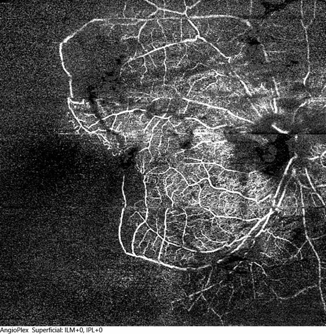
- optical coherence tomography (OCT), central retinal vein occlusion (CRVO), ischemic CRVO, ultra-wide field imaging
- Photographer
- Eduardo Torres-Porras, Provissia, Laser y Ultrasonido Ocular
- Imaging device
-
Optical coherence tomography system
Cirrus 600, Carl Zeiss - Description
- 12x12 mm OCT angiogram of the right eye of a 36-year-old male who had an ischemic CRVO following a hypertensive emergency secondary to consumption of high doses of cocaine. Areas of non perfusion are found within the posterior pole and ischemia extends temporally to the mid-periphery.


")


")
")
")