-
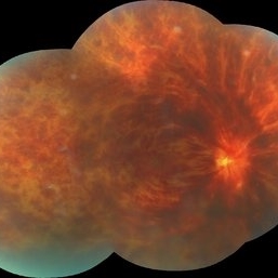
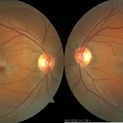
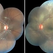
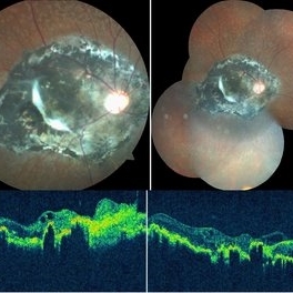
 CRVO
CRVO
Nov 26 2020 by Priya Rasipuram Chandrasekaran, MBBS, DO, DNB, FRCS
A 44-year-old male patient presented with no underlying systemic illness presented with this picture showing extensive scattered superficial and deep retinal hemorrhages to confluent retinal hemorrhages extending to all the quadrants associated with marked dilatation and tortuosity of vessels and associated with optic disc edema, macular edema and retinal thickening giving the appearance of blood and thunder retina.
Condition/keywords: central retinal vein occlusion (CRVO)
-
 Central Retinal Vein Occlusion
Nov 26 2020 by Priya Rasipuram Chandrasekaran, MBBS, DO, DNB, FRCS
A 44-year-old male patient presented with no underlying systemic illness presented with this picture showing extensive scattered superficial and deep retinal hemorrhages to confluent retinal hemorrhages extending to all the quadrants associated with marked dilatation and tortuosity of vessels and associated with optic disc edema, macular edema and retinal thickening giving the appearance of blood and thunder retina.
Condition/keywords: central retinal vein occlusion (CRVO)
-
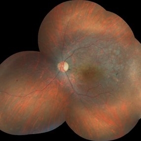
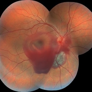
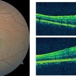
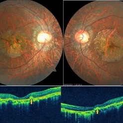
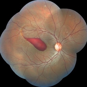 Preretinal Hemorrhage Extending into the Macula
Nov 26 2020 by Priya Rasipuram Chandrasekaran, MBBS, DO, DNB, FRCS
A 26-year-old male with no h/o trauma or underlying systemic disease presented with the complaint of central scotoma in the right eye since 1 month and fundus examination showed preretinal hemorrhage in the supero-temporal quadrant extending into the macular area and OCT macula showing premacular hemorrhage.
Condition/keywords: preretinal hemorrhage
-
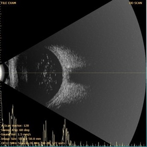
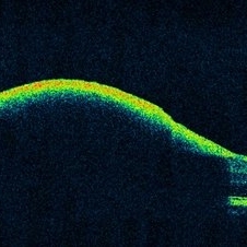 OCT Showing Premacular Hemorrhage
Nov 26 2020 by Priya Rasipuram Chandrasekaran, MBBS, DO, DNB, FRCS
A 26-year-old male with no h/o trauma or underlying systemic disease presented with the complaint of central scotoma in the right eye since 1 month and fundus examination showed preretinal hemorrhage in the supero-temporal quadrant extending into the macular area and OCT macula showing premacular hemorrhage.
Condition/keywords: premacular hemorrhage
-
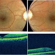
 ERM
ERM
Nov 26 2020 by Priya Rasipuram Chandrasekaran, MBBS, DO, DNB, FRCS
A 58-year-old female presented with distortion of images 1 month following cataract surgery in the right eye and fundus examination showed epiretinal membrane extending from the disc to the macula and OCT macula showing epiretinal membrane with disorganization of the foveal architecture.
Condition/keywords: epiretinal membrane (ERM)
-
 OCT Showing ERM
Nov 26 2020 by Priya Rasipuram Chandrasekaran, MBBS, DO, DNB, FRCS
A 58-year-old female presented with distortion of images 1 month following cataract surgery in the right eye and fundus examination showed epiretinal membrane extending from the disc to the macula and OCT macula showing epiretinal membrane with disorganization of the foveal architecture.
Condition/keywords: epiretinal membrane (ERM)
-
 RD With Macular Hole
Nov 27 2020 by Priya Rasipuram Chandrasekaran, MBBS, DO, DNB, FRCS
A 42-year-old female presented with sudden decrease in vision in the right eye and fundus examination showed bullous retinal detachment.
Condition/keywords: acute retinal detachment
-
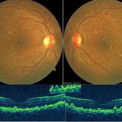
 Macular-BRVO
Nov 27 2020 by Priya Rasipuram Chandrasekaran, MBBS, DO, DNB, FRCS
A 48-year-old female with no underlying known systemic illness presented with decreased vision in the left eye and fundus showed macular branch retinal vein occlusion.
Condition/keywords: macular branch retinal vein occlusion (BRVO)
-
 Idiopathic Parafoveal Telengiectasia - Type 2
Nov 27 2020 by Priya Rasipuram Chandrasekaran, MBBS, DO, DNB, FRCS
This is the fundus photo of a 61-year-old male presenting with bilateral gradual loss of vision since 6 months. Bilateral fundus photo shows parafoveal greying with early crystals and retinal pigment hyperplasia around a dilated venule suggestive of type 2 idiopathic parafoveal telangiectasia.
Condition/keywords: parafoveal telangiectasia
-
 Central-RP
Nov 28 2020 by Priya Rasipuram Chandrasekaran, MBBS, DO, DNB, FRCS
Central or inverse retinitis pigmentosa.
Condition/keywords: retinitis pigmentosa
-
 Trio of Retinal Hemorrhages
Dec 8 2020 by Priya Rasipuram Chandrasekaran, MBBS, DO, DNB, FRCS
This is the fundus photo of a 29-year-old following blunt trauma showing hemorrhages in all the three layers of the retina (vitreous hemorrhage, subhyaloid hemorrhage and subretinal hemorrhage)
Condition/keywords: blunt trauma, retinal hemorrhage
-
 Ultrasound B-Scan of asteroid hyalosis
Dec 8 2020 by Priya Rasipuram Chandrasekaran, MBBS, DO, DNB, FRCS
Ultrasound B-Scan shows discrete mobile point / dot like bright echoes in the vitreous cavity with an acoustic clear zone between the dot echoes and the posterior globe wall with no posterior acoustic shadowing. The corresponding A scan shows high spikes. OCT macula confirms the same.
Condition/keywords: asteroid hyalosis
-
 TRD with Macular Hole
Dec 8 2020 by Priya Rasipuram Chandrasekaran, MBBS, DO, DNB, FRCS
Horizontal 5 line raster scan through the macula shows full thickness macular hole along with separation of neuro sensory retina from the retinal pigment epithelium.
Condition/keywords: acute retinal detachment, traumatic macular hole
-
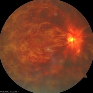
 Valslava Retinopathy
Jan 15 2021 by Priya Rasipuram Chandrasekaran, MBBS, DO, DNB, FRCS
This is the fundus photo and red free montage showing preretinal hemorrhage of the left eye along the superior retina, abutting the disc margin and extending as far as the macula. There are few scattered flame shaped hemorrhages superiorly, nasally and inferiorly with a central white spot mimicking Roth spots.
Condition/keywords: valsalva retinopathy
-
 CHRPE
CHRPE
Jan 15 2021 by Priya Rasipuram Chandrasekaran, MBBS, DO, DNB, FRCS
This is the fundus photo and fundus photo montage of the left eye of a 25-year-old male showing flat, solitary, round, greyish pigmented lesion situated AT THE equator with a scalloped margin. Vessels overlying the lesion are normal and there is a clear demarcation line between this and normal retina. The margins are hypopigmented with few hypopigmented lacunae inside.
Condition/keywords: congenital hypertrophy of the retinal pigment epithelium (CHRPE)
-
 Choroidal Rupture
Apr 7 2021 by Priya Rasipuram Chandrasekaran, MBBS, DO, DNB, FRCS
Choroidal rupture
Condition/keywords: choroidal rupture
-
 Choroidal Rupture
Apr 7 2021 by Priya Rasipuram Chandrasekaran, MBBS, DO, DNB, FRCS
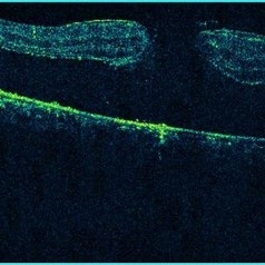
The fundus photo of a 24-year-old male shows crescent shaped choroidal rupture away from fovea and concentric to the optic disc following cricket ball injury. The corresponding optical coherence tomography shows disruption of the choriocapillaris, retinal pigment epithelium and Bruch’s membrane while the neurosensory retina remains intact. The fovea is not involved.
Condition/keywords: choroidal rupture
-
 Macular Scar
Apr 7 2021 by Priya Rasipuram Chandrasekaran, MBBS, DO, DNB, FRCS
The fundus photo shows macular scar with epiretinal membrane in a 22-year-old female presenting with 20/80 N10 vision. The corresponding optical coherence tomography (vertical and horizontal raster scan) shows fibroglial proliferation with epiretinal membrane and traction at the fovea.
Condition/keywords: macular scar, scar
-
 Spontaneous Reattachment of Retinal Detachment
Apr 26 2021 by Priya Rasipuram Chandrasekaran, MBBS, DO, DNB, FRCS
This is the fundus photo montage and red free montage of a 27-year-old male showing pigmentary changes and atrophic changes in the inferior retina involving the macula. This has sharply demarcated margins and a convex border with subretinal bands suggestive of spontaneous reattachement of retinal detachment.
Condition/keywords: re-attached retinal detachment (RRD), spontaneous retinal reattachment
-
 PRP Marks
Apr 26 2021 by Priya Rasipuram Chandrasekaran, MBBS, DO, DNB, FRCS
This is the fundus photo montage of both eyes of a patient showing pan retinal photocoagulation marks. Theses marks can be confused with gyrate atrophy, cobble stone degeneration and myopic degeneration.
Condition/keywords: pan-retinal photocoagulation (PRP)
-
 Commotio Retinae
Apr 27 2021 by Priya Rasipuram Chandrasekaran, MBBS, DO, DNB, FRCS
This is the fundus photo montage of a 11-year-old boy showing extensive commotio retinae evidenced by the absence of greyish opacification of the retina with the absence of the usual red reflex following blunt trauma to the left eye. There is no pseudo cherry red spot. The right eye has been added for comparison.
Condition/keywords: commotio retinae
-
 Welders Maculopathy
Apr 27 2021 by Priya Rasipuram Chandrasekaran, MBBS, DO, DNB, FRCS
Fundus photo of both eyes showing absent foveal reflex and orange red discolouration surrounded by a pigmentary halo. The corresponding OCT image of the macula shows outer retinal hole extending from the inner layer of retinal pigment epithelium to the external limiting membrane which inturn corresponds to IS/OS junction.
Condition/keywords: Welder's maculopathy
-
 Fundus Albipunctatus
Apr 27 2021 by Priya Rasipuram Chandrasekaran, MBBS, DO, DNB, FRCS
This is the fundus photo montage of a 23-year-old male demonstrating whitish-yellow spots all over the fundus sparing the fovea at the level of retinal pigment epithelium. This belongs to the group of congenital stationary night blindness with flecks in the retina.
Condition/keywords: fleck retinopathy
-
 Retinal Blood Vessels in Retinochoroidal (RC) Coloboma
May 4 2021 by Priya Rasipuram Chandrasekaran, MBBS, DO, DNB, FRCS
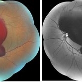
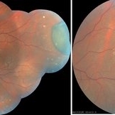
This is the fundus photo of a 10-year-old girl showing RC coloboma along the infero nasal retina and involving the disc. This belongs to grade 4 of Ida Mann’s classification and grade 5 of Lingam Gopal’s classification of RC coloboma. The optic disc has no cup and BV for superior fundus emanates from superior part of optic disc and that for inferior fundus in the colobomatous area from multiple points. The blood vessels are discontinuous and are cork screw shaped.
Condition/keywords: chorioretinal coloboma
-
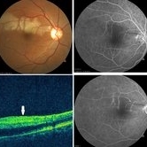 Branch Retinal Artery Occlusion
May 4 2021 by Priya Rasipuram Chandrasekaran, MBBS, DO, DNB, FRCS
This is the fundus photo of a 52-year-old male taken within 6 hours and after 24 hours of sudden onset of inferior field loss. The photo shows prominence of retinal edema in the region of arterial occlusion as time passes by. The optical coherence tomogram scan taken vertically through the normal and the involved area shows thickening and hyper reflectivity of retinal nerve fiber layer and decreased reflectivity of the retinal layers beneath it (white arrow). Fundus fluorescein angiography shows complete non-filling of the artery in the early phase with slow filling in the late phase and highlighting the embolus.
Condition/keywords: branch retinal artery occlusion (BRAO)
-
 Chorioretinitis Sclopetaria
May 4 2021 by Priya Rasipuram Chandrasekaran, MBBS, DO, DNB, FRCS
This fundus photo and montage shows pigmentary changes with fibroglial proliferation of the disc and macula in a 36-year-old male following injury with an iron chain. This is usually following a high velocity non-penetrating missile or blast injury categorized as coup injury and can be both direct or indirect. The layers affected are the highly inelastic Bruch’s membrane with choriocapillaris and retinal pigment epithelium in contrast to the highly elastic retina and sclera. The high impact injury causes full thickness defect in the retina, Bruch’s membrane and choroid leading to retraction of the retina and choroid, leaving the intact bare sclera behind. Pathology included defects in the Bruch’s membrane and choroid, and extensive photoreceptor loss with hyperplasia of retinal pigment epithelium. Over the weeks, loose fibrous tissue gets replaced by dense connective tissue leading to scarring between retina and choroid as seen in our patient. The background shows fundus albipunctatus.
Condition/keywords: chorioretinitis sclopetaria
-
 Central Areolar Choroidal Dystrophy
May 4 2021 by Priya Rasipuram Chandrasekaran, MBBS, DO, DNB, FRCS
Fundus photo of a 34-year-old male showing bilaterally symmetrical atrophy of retinal pigment epithelium (RPE) and choriocapillaris involving the fovea and highlighting the underlying large choroidal vessels. OCT macula shows atrophy of the outer retinal layers up to the external limiting membrane along with thinning of RPE and Bruch's membrane complex. Rosette - like hyperreflective structures causing retinal elevation at the border of atrophic area (yellow arrows) are seen categorizing this into stage 4 disease.
Condition/keywords: central areolar choroidal dystrophy (CACD)
-
 Myelinated Nerve Fiber
May 5 2021 by Priya Rasipuram Chandrasekaran, MBBS, DO, DNB, FRCS
A 31-year-old male presented with a decreased vision of 20/125 N24 with -6.50 DS/-3.50 cyl 90 in the left eye. Fundus examination revealed peripapillary MNF progressing superiorly, obscuring disc and vessels and sparing the macula. OCT of ONH showed hyper reflective NFL and an abrupt ending of RPE and inner retinal layers (IRL) with underlying shadowing at the beginning of hyper reflectivity. The absence of photoreceptor integrity line (PIL) in the macula is believed to cause refractory amblyopia in such patients.
Condition/keywords: myelinated nerve fibers
-
 Ozurdex Implant
May 6 2021 by Priya Rasipuram Chandrasekaran, MBBS, DO, DNB, FRCS
This is the fundus photo of a patient with Ozurdex implant containing 0.7 m of dexamethasone given for gross cystoid macular edema in multifocal choroidtitis.
Condition/keywords: dexamethasone implant
-
 Cuticular Drusen
Jun 13 2021 by Priya Rasipuram Chandrasekaran, MBBS, DO, DNB, FRCS
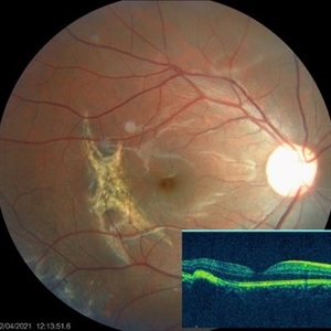
This is the fundus photo showing numerous yellow, small, hard drusen distributed throughout the retina. The corresponding OCT shows numerous elevated lesions underneath the RPE causing RPE elevations and arranged in a saw-tooth manner. Macular complications include acquired vitelliform lesion, choroidal neovascular membrane and geographic atrophy which are common after 60 years of age. It is usually associated with mutations in complement factor H. Basal laminar drusen, diffuse drusen and early adult onset grouped drusen are other alternative names. The differential diagnosis includes autosomal dominant drusen, pattern macular dystrophy, Sorsby macular drusen, mitochondrial macular dystrophy and so on.
Condition/keywords: cuticular drusen
-
 CRVO
CRVO
Nov 26 2020 by Priya Rasipuram Chandrasekaran, MBBS, DO, DNB, FRCS
A 44-year-old male patient presented with no underlying systemic illness presented with this picture showing extensive scattered superficial and deep retinal haemorrhages to confluent retinal hemorrhages extending to all the quadrants associated with marked dilatation and tortuosity of vessels and associated with optic disc edema, macular edema and retinal thickening giving the appearance of blood and thunder retina.
Condition/keywords: central retinal vein occlusion (CRVO)
-
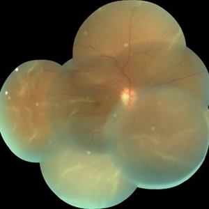
 Central Retinal Vein Occlusion
Nov 26 2020 by Priya Rasipuram Chandrasekaran, MBBS, DO, DNB, FRCS
A 44-year-old male patient presented with no underlying systemic illness presented with this picture showing extensive scattered superficial and deep retinal hemorrhages to confluent retinal hemorrhages extending to all the quadrants associated with marked dilatation and tortuosity of vessels and associated with optic disc edema, macular edema and retinal thickening giving the appearance of blood and thunder retina.
Condition/keywords: central retinal vein occlusion (CRVO)
A project from the American Society of Retina Specialists