 Initializing download.
-
 By Priya Rasipuram Chandrasekaran, MBBS, DO, DNB, FRCS
Lotus eye hospital
Co-author(s): Lotus eye hospital, Salem - Uploaded on Jan 15, 2021.
- Last modified by Caroline Bozell on Jan 19, 2021.
- Rating
- Appears in
- Miscellaneous
- Condition/keywords
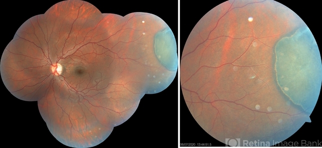
- congenital hypertrophy of the retinal pigment epithelium (CHRPE)
- Imaging device
- Fundus camera
- Description
- This is the fundus photo and fundus photo montage of the left eye of a 25-year-old male showing flat, solitary, round, greyish pigmented lesion situated AT THE equator with a scalloped margin. Vessels overlying the lesion are normal and there is a clear demarcation line between this and normal retina. The margins are hypopigmented with few hypopigmented lacunae inside.


")
---thumb.jpg/image-square;max$79,0.ImageHandler "Macular CHRPE")
")
")
")


