 Initializing download.
-
 By Priya Rasipuram Chandrasekaran, MBBS, DO, DNB, FRCS
Lotus eye hospital
Co-author(s): Lotus eye hospital, Salem - Uploaded on Jun 13, 2021.
- Last modified by Caroline Bozell on Jun 15, 2021.
- Rating
- Appears in
- Miscellaneous
- Condition/keywords
- cuticular drusen
- Imaging device
- Fundus camera
- Description
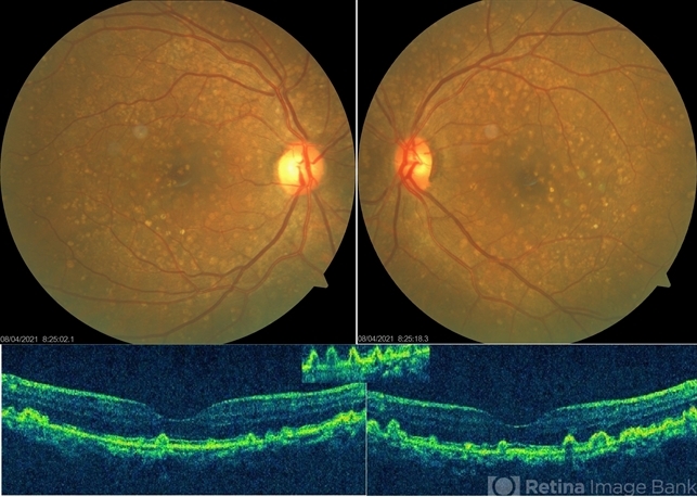
- This is the fundus photo showing numerous yellow, small, hard drusen distributed throughout the retina. The corresponding OCT shows numerous elevated lesions underneath the RPE causing RPE elevations and arranged in a saw-tooth manner. Macular complications include acquired vitelliform lesion, choroidal neovascular membrane and geographic atrophy which are common after 60 years of age. It is usually associated with mutations in complement factor H. Basal laminar drusen, diffuse drusen and early adult onset grouped drusen are other alternative names. The differential diagnosis includes autosomal dominant drusen, pattern macular dystrophy, Sorsby macular drusen, mitochondrial macular dystrophy and so on.
 drusen - colour RE")
 drusen - colour LE")
 drusen - FA 1")
 drusen - FA 2")


