 Initializing download.
-
 By Priya Rasipuram Chandrasekaran, MBBS, DO, DNB, FRCS
Lotus eye hospital
Co-author(s): Lotus eye hospital, Salem - Uploaded on May 4, 2021.
- Last modified by Caroline Bozell on May 4, 2021.
- Rating
- Appears in
- Miscellaneous
- Condition/keywords
- branch retinal artery occlusion (BRAO)
- Imaging device
- Fundus camera
- Description
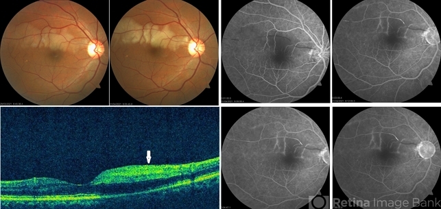
- This is the fundus photo of a 52-year-old male taken within 6 hours and after 24 hours of sudden onset of inferior field loss. The photo shows prominence of retinal edema in the region of arterial occlusion as time passes by. The optical coherence tomogram scan taken vertically through the normal and the involved area shows thickening and hyper reflectivity of retinal nerve fiber layer and decreased reflectivity of the retinal layers beneath it (white arrow). Fundus fluorescein angiography shows complete non-filling of the artery in the early phase with slow filling in the late phase and highlighting the embolus.


---thumb.jpg/image-square;max$79,0.ImageHandler "Branch Retinal Artery Occlusion")
