Search results (0 results)
-
 Sunset Glow Fundus
Oct 14 2024 by César Adrián Gómez Valdivia, MD
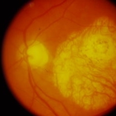
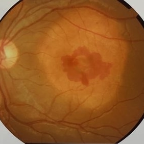
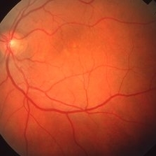
"Sunset Glow Fundus” found in a 65 year-old male patient diagnosed with Voght-Koyanagi-Harada disease, convalescent stage. Choroidal depigmentation occurs several months after the uveitic stage, leading to a pale disc with a bright red-orange choroid.
Photographer: @eyemissu2
Imaging device: TOPCON TRC-50DX
Condition/keywords: Sunset, Sunset Glow Fundus
-
 Sunset Glow Fundus
Oct 14 2024 by César Adrián Gómez Valdivia, MD
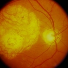
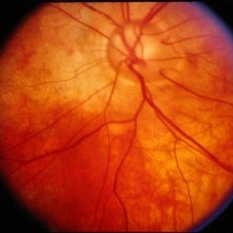
"Sunset Glow Fundus” found in a 65 year-old male patient diagnosed with Voght-Koyanagi-Harada disease, convalescent stage. Choroidal depigmentation occurs several months after the uveitic stage, leading to a pale disc with a bright red-orange choroid.
Photographer: @eyemissu2
Imaging device: TOPCON TRC-50DX
Condition/keywords: sunset, Sunset Glow Fundus
-
 Sunset Glow Fundus
Oct 14 2024 by César Adrián Gómez Valdivia, MD
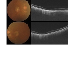
"Sunset Glow Fundus” found in a 65 year-old male patient diagnosed with Voght-Koyanagi-Harada disease, convalescent stage. Choroidal depigmentation occurs several months after the uveitic stage, leading to a pale disc with a bright red-orange choroid.
Photographer: @eyemissu2
Imaging device: California ICG OPTOS
Condition/keywords: sunset, Sunset Glow Fundus, vkh
-
 Sunset Glow Fundus
Oct 14 2024 by César Adrián Gómez Valdivia, MD
“Sunset Glow Fundus” found in a 65 year-old male patient diagnosed with Voght-Koyanagi-Harada disease, convalescent stage. Choroidal depigmentation occurs several months after the uveitic stage, leading to a pale disc with a bright red-orange choroid.
Photographer: @eyemissu2
Imaging device: OPTOS
Condition/keywords: sunset, Sunset Glow Fundus, VKH
-
 Choroidal Osteoma
Sep 12 2023 by Ben Serar
Fundus photograph of the LE showing an irregular, yellow-white, juxtapapillary, choroidal lesion with well-defined geographic borders; with diffuse and mottled depigmentation of the overlying pigment epithelium; and multiple small vascular networks on the tumor surface.
Condition/keywords: choroidal osteoma
-
 Choroidal Osteoma
Sep 12 2023 by Ben Serar
Fundus photograph of the RE showing an irregular, yellow-white, juxtapapillary, choroidal lesion with well-defined geographic borders; with diffuse and mottled depigmentation of the overlying pigment epithelium; and multiple small vascular networks on the tumor surface.
Condition/keywords: choroidal osteoma
-
 Central Areolar Choroidal Dystrophy
Oct 30 2020 by Mihir Trivedi
Fundus photo of a 43-year-old female with gradual onset diminution of vision in both eyes since 2-3 years. BCVA in OU was 3/60. She was diagnosed to have central areolar choroidal dystrophy(CACD). Central areolar choroidal dystrophy (CACD) is a rare inherited disease, which causes progressive profound loss of vision in patients during their fourth decade. It is characterized by atrophy of retinal pigment epithelium, photoreceptors and choriocapillaris. IT is a progressive macular dystrophy characterized by subtle, mottled depigmentation in the posterior pole in the early stages. The depigmentation area gradually enlarges until an oval or round surface of atrophy of the retinal pigmentary epithelium and choriocapillaris is formed. Drusen or flecks are absent in a typical presentation.
Photographer: Mr Ganesh Naidu
Imaging device: TOPCON DRI Triton
Condition/keywords: central areolar choroidal dystrophy (CACD)
-
 Enucleated Eye with Retinal Atrophy
May 18 2020 by McGill University Health Centre
This image illustrates marked retinal atrophy with several bone-spicule-shaped pigment deposits in the peripheral retina. The macular area is preserved but has a rim of depigmentation. Note the thin blood vessels and the pallor of the optic nerve.
Condition/keywords: atrophy, enucleation
-
 Choroidal Osteoma With Active CNVM
Apr 10 2020 by Dipak Nag, MBBS, FCPS, MSc, FRF
A 12-year-old boy visited our clinic for sudden, painless blurring of vision and metamorphopsia in left eye that he noticed 7 days back. His BCVA was 6/60 in left eye. Anterior segment examination was unremarkable. On fundus examination of his left eye showed a yellow-white lesion at the macula with well-defined geographic border and diffuse and mottled depigmentation of the overlying pigment epithelium, of which an elevated gray-green areas at the center with subretinal hemorrhage around. The right eye was found normal.
Photographer: Mr. Shamsuddin
Condition/keywords: choroidal neovascular membrane (CNVM)
-
 Vogt-Koyanagi-Harada Disease
Dec 13 2019 by McGill University Health Centre
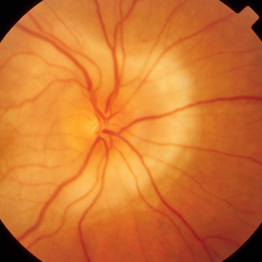
Fundus photograph of an 45-year-old Asian descendant woman with poliosis and periocular vitiligo. Fundoscopy reveals a large area of depigmentation of the fundus and a retinal detachment .
Photographer: Miguel N. Burnier, McGill University Health Center-McGill University Ocular Pathology & Translational Research Laboratory
Imaging device: Fundoscopy
Condition/keywords: depigmentation of the fundus, Vogt-Koyanagi-Harada
-
 Vogt-Koyanagi-Harada Disease
Dec 13 2019 by McGill University Health Centre
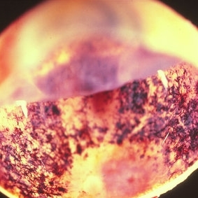
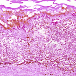
An 45-year-old Asian descendant woman with poliosis and periocular vitiligo. Histopathology shows thickening of the choroid and chronic granulomatous choroiditis involving the choriocapillaris, typical of VKH.
Photographer: Miguel N. Burnier, McGill University Health Center-McGill University Ocular Pathology & Translational Research Laboratory
Imaging device: Zeiss
Condition/keywords: depigmentation of the fundus, histopathology, Vogt-Koyanagi-Harada
-
 Central Serous Retinopathy
Mar 26 2019 by Gary R. Cook, MD, FACS
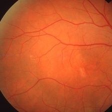
45-year-old white male with history of bilateral CSR; Color fundus photo showing an area of RPE depigmentation in temporal macula; VA = 20/200.
Imaging device: Topcon VT-50
Condition/keywords: central serous retinopathy (CSR)
-
 Central Serous Retinopathy
Mar 26 2019 by Gary R. Cook, MD, FACS
45-year-old white male with a history of bilateral CSR showing subtle RPE depigmentation in macula and inferiorly in his symptomatic OS; VA = 20/30.
Imaging device: Topcon VT-50
Condition/keywords: central serous retinopathy (CSR)
-
 Bilateral Central Serous Retinopathy
Mar 26 2019 by Gary R. Cook, MD, FACS
Asymptomatic left eye of a 37-year-old white male with a history of previous CSR OS showing some focal RPE depigmentation perifoveally and subretinic deposits temporally; no NSRD is present; VA = 20/15+3.
Imaging device: Topcon VT-50
Condition/keywords: central serous retinopathy (CSR), resolved subretinal fluid, retinal pigment epithelium (RPE) changes
-
 Choroideremia Carrier Female
Oct 25 2018 by INDU V P, MBBS, MS
Fundus photograph of a 26-year-old carrier female whose father has choroideremia. Choroideremia is a rare genetic disease with x-linked recessive inheritance. Affected males have nightblindness with progressive loss of peripheral vision. Carrier females are asymptomatic but may have patchy depigmentation of retinal pigment epithelium
Condition/keywords: choroideremia carrier, x linked recessive
-
 Choroideremia
Oct 25 2018 by INDU V P, MBBS, MS
Fundus photograph of a 26-year-old carrier female whose father has choroideremia. Choroideremia is a rare genetic disease with x-linked recessive inheritance. Affected males have nightblindness with progressive loss of peripheral vision. Carrier females are asymptomatic but may have patchy depigmentation of retinal pigment epithelium
Condition/keywords: choroideremia carrier, x linked recessive
-
 Retinitis Pigmentosa
Aug 25 2015 by René Hernán Parada Vásquez
Fundus photograph of both eyes of a 38-year-old female with retinitis pigmentosa, bone spicule-shaped pigment deposits are present in the mid periphery, and macula with a peripheral ring of depigmentation.
Photographer: Parada René, ESO, Guatemala.
Imaging device: Canon CR-2
Condition/keywords: bilateral pigmentary retinopathy, retinitis pigmentosa, retinitis pigmentosa (RP) dystrophy
-
 Linear Nevus Sebaceous Syndrome
Feb 20 2015 by H. Michael Lambert, MD
Linear nevus sebaceous syndrome color photo with choroidal depigmentation.
Condition/keywords: choroidal depigmentation, linear nevus sebaceous syndrome
-
---thumb.jpg/image-square;max$300,300.ImageHandler) Flecked Retina
Flecked Retina
Apr 4 2014 by H. Michael Lambert, MD
Age 9, flecked retina, 20/20 OU.
Condition/keywords: depigmentation
-
---thumb.jpg/image-square;max$300,300.ImageHandler) Flecked Retina
Flecked Retina
Apr 4 2014 by H. Michael Lambert, MD
Age 9, flecked retina, 20/20 OU.
Condition/keywords: depigmentation
-
---thumb.jpg/image-square;max$300,300.ImageHandler) AMPPE
AMPPE
Aug 13 2013 by From the Collections of Thomas M. Aaberg, MD and Thomas M. Aaberg Jr., MD
Chronic depigmentation.
Condition/keywords: acute posterior multifocal placoid pigment epitheliopathy (APMPPE), depigmentation
-
---thumb.jpg/image-square;max$300,300.ImageHandler) AMPPE
AMPPE
Aug 13 2013 by From the Collections of Thomas M. Aaberg, MD and Thomas M. Aaberg Jr., MD
Chronic depigmentation.
Condition/keywords: acute posterior multifocal placoid pigment epitheliopathy (APMPPE), depigmentation
-
---thumb.jpg/image-square;max$300,300.ImageHandler) AMPPE
AMPPE
Aug 13 2013 by From the Collections of Thomas M. Aaberg, MD and Thomas M. Aaberg Jr., MD
Chronic depigmentation.
Condition/keywords: acute posterior multifocal placoid pigment epitheliopathy (APMPPE), depigmentation
-
---thumb.jpg/image-square;max$300,300.ImageHandler) AMPPE
AMPPE
Aug 13 2013 by From the Collections of Thomas M. Aaberg, MD and Thomas M. Aaberg Jr., MD
Chronic depigmentation, FA.
Condition/keywords: acute posterior multifocal placoid pigment epitheliopathy (APMPPE), depigmentation
-
---thumb.jpg/image-square;max$300,300.ImageHandler) AMPPE
AMPPE
Aug 13 2013 by From the Collections of Thomas M. Aaberg, MD and Thomas M. Aaberg Jr., MD
Chronic depigmentation.
Condition/keywords: acute posterior multifocal placoid pigment epitheliopathy (APMPPE), depigmentation
 Loading…