Search results (32 results)
-
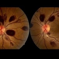
 Prepapillary Vascular Loop
Jul 4 2025 by KANWALJEET HARJOT MADAN, M.S. (Ophthalmology); FAICO (Vitreous - Retina)
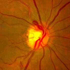
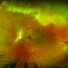
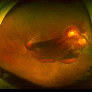
This is the fundus picture of right eye of a young 32 years female depicting pre papillary vascular loop. A prepapillary vascular loop is a congenital anomaly of the optic disc that presents as an elevated and twisted bundle of vessels projecting into the vitreous cavity. It is a benign condition, usually unilateral but can be bilateral. It is asymptomatic and discovered during routine eye examination. This anomaly can sometimes cause complications like branch retinal artery occlusion, vitreous hemorrhage, or sub retinal hemorrhage.
Photographer: Dr. Kanwaljeet Harjot Madan, Thind Eye Hospital, Jalandhar City (Punjab) INDIA.
Imaging device: Zeiss Fundus Camera
Condition/keywords: branch retinal artery occlusion (BRAO), optic disc, Prepapillary Vascular Loop, SUB RETINAL HEMORRHAGE, Vitreous hemorrhage
-
 Choroidal Fracture
Oct 27 2024 by César Adrián Gómez Valdivia, MD
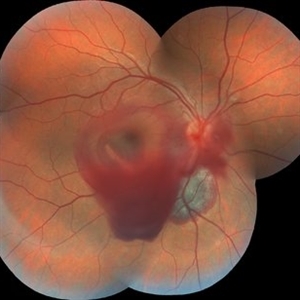
Fundus photograph of a traumatic choroidal fracture & extra-macular sub-retinal hemorrhage.
Photographer: @eyemissu2
Imaging device: TOPCON TRC-50DX
Condition/keywords: Choroidal Fracture
-
 Dislocated Lens, Posterior OD
Jan 26 2024 by Corey Grant
OPTOS California photo presents a 71 year old male patient with a dislocated lens, posterior in the right eye. Presented on 1/26/24 with posteriorly dislocated SN60WF with a Soemmerring ring. Associated retinal hemorrhage within retinoschisis as well. This will result in a PPV/IOL exchange/SFIOL/STK for the right eye.
Photographer: Corey Grant, Ophthalmic Imager, Retina Specialist of Michigan
Imaging device: OPTOS California
Condition/keywords: color photo, IOL, OD, Optos, OPTOS CALIFORNIA, pars plana vitrectomy (PPV), retina
-
 PEHCR (Peripheral Exudative Hemorrhagic Chorioretinopathy)
May 12 2023 by Niloofar Piri, MD
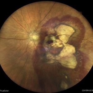
Ultrawide fundus photograph of the left eye demonstrating extensive peripheral hemorrhagic exudative detachment in a 79 yo Caucasian female with prior history of non-exudative AMD. Recent diagnosis of Acute myeloid leukemia with low platelet count which might have contributed to the above presentatuon. Please note the temporal subretinal hemorrhage as well as RPE atrophy and hyperplasia in the macula.
Photographer: Rocio Bentivegna, MD, Saint Louis University; Jessica Maddox, COA, Saint Louis University
Condition/keywords: peripheral exudative hemorrhagic chorioretinopathy (PEHCR)
-
 Subretinal Hemorrhage
Feb 28 2023 by Akansha Sharma
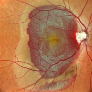
Color fundus photograph of an 84-year old male with subretinal hemorrhage associated with areas of scarring.
Photographer: Dr. Urmil Shah, Dr. Denish Patel, Dr. Akansha Sharma, Bharati Eye Hospital, Ahmedabad, Gujarat
Condition/keywords: choroidal neovascularization (CNV), subretinal hemorrhage
-
 Lady in a dress
Feb 9 2023 by Shelby Helton
Fluorescein Angiography on a 67-year-old male with significant RPE changes secondary to a severe subretinal hemorrhage that required a vitrectomy with subretinal TPA in 2013.
Photographer: Shelby Helton
Imaging device: Heidelberg Spectralis
Condition/keywords: wet age-related macular degeneration (wet AMD)
-
 High risk Proliferative Diabetic Retinopathy treated with Pan Retinal Photocoagulation
Nov 5 2022 by Somnath Chakraborty, MD
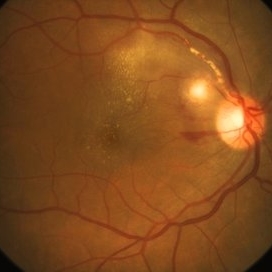
A Fundus Photo Montage of 43 year old Asian Male with Type 2 Diabetes Mellitus since 7 years who presented with sudden onset diminition of vision in his Left eye. BCVA OS was 20/200. He was diagnosed to have Pre retinal bleed due to Proliferative Diabetic Retinopathy and was treated with Pan Retinal Photocoagulation. This image shows a large neo-cascular frond at the disc and superior to it with Pre-retinal bleed and Fresh laser marks along
Photographer: Pulak Roy
Condition/keywords: diabetic blindness, diabetic retinopathy vitrectomy study (DRVS), fresh laser burns, laser photocoagulation, preretinal hemorrhage, proliferative diabetic retinopathy (PDR)
-
 Choroidal Melanoma
Nov 3 2022 by pedro fernandes souza neto
Transillumination of Enucleation specimen of Choroidal Melanoma: anterior chamber is closed. Total secondary retinal detachment with subretinal serous fluid and some subretinal hemorrhages are present.
Photographer: Eduardo Marback, Federal University of Bahia, Brazil
Condition/keywords: enucleation, melanoma
-
 Subretinal BSS and air
Apr 12 2022 by Nassim Alejandro Abreu Arbaje, MD
67 year old female who presented with complaints of 5 days of decreased vision of her left eye. She underwent PPV + BSS and Air injection in the subretinal space
Photographer: Nassim Abreu, Dr. Elias Santana Hospital
Imaging device: Ngenuity 3D system screenshot
Condition/keywords: subretinal hemorrhage
-
 Subretinal Bleed
Jul 12 2022 by Akansha Sharma
73 year old diabetic and hypertensive female presented with sub-retinal hemorrhage for which she was operated with pars-plana vitrectomy with intra-vitreal anti-VEGF
Photographer: Dr. Akansha Sharma-Retina Foundation, Ahmedabad
Condition/keywords: subretinal hemorrhage, subretinal blood
-
 Cat Scratch Disease
Mar 29 2021 by Gabriel Costa Andrade, PhD
Fundus photograph of an 36-year-old woman with a macular vasculitis, pre retinal hemorrhage and exudation due to Bartonella henselae infection.
Photographer: Gabriel Andrade
Condition/keywords: cat scratch retinitis
-
 Massive Commotio Retinae
Oct 20 2020 by Veronika Yehezkeli
Fundus photograph of a 24-year-old male, made after blunt trauma with a plastic bottle. Note massive commotio retinae and preretinal hemorrhages in the contralateral to trauma area.
Photographer: Veronika Yehezkeli, Meir medical center, Israel
Condition/keywords: blunt trauma, commotio retinae, preretinal hemorrhage, trauma
-
 Trio of Retinal Hemorrhages
Dec 8 2020 by Priya Rasipuram Chandrasekaran, MBBS, DO, DNB, FRCS
This is the fundus photo of a 29-year-old following blunt trauma showing hemorrhages in all the three layers of the retina (vitreous hemorrhage, subhyaloid hemorrhage and subretinal hemorrhage)
Condition/keywords: blunt trauma, retinal hemorrhage
-
 CMV Retinitis with Frosted Branch Angiitis
Sep 23 2020 by Nimesh A. Patel, MD, FASRS
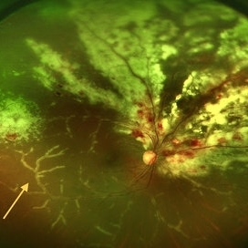
Fundus photo showing peri-vascular inflammation of both arteries and veins with translucent exudation (yellow arrow). Superior nasally, there is classic retinal whitening with retinal hemorrhages superior. This patient was found to have a low CD4 count and a diagnosis of AIDS was made.
Condition/keywords: cytomegalovirus (CMV), HIV, uveitis
-
 Blunt Ocular Trauma Due to Firework Injury
Jun 9 2020 by Brittany Rota
Ultra- widefield pseudocolor image of an 18-year-old male with blunt ocular trauma in the right eye due to a firework injury. The patient presented with commotio retinae (sclopteria), an acute vitreous hemorrhage, choroidal rupture, and a subretinal hemorrhage. The referring physician performed surgery on the lateral rectus muscle which was macerated but not severed, and several orbital fibrous foreign bodies were removed from the posterior orbit. The globe was intact. There is no evidence of retinal tear in the region of sclopetaria; however, there is complete necrosis of the temporal peripheral choroid and retina. The vitreous hemorrhage was slowly clearing on his exam 6-9-2020. The patient is developing subretinal fibrosis. The physician is concerned about the choroidal rupture that is visible through the submacular hemorrhage. There is one rupture that appears to course directly under the fovea. The physician states that if this is the case, his vision most likely will be 20/200 or worse. His vision was hand motion in all fields except nasally, which he was unable to see hand motion at his visit on 6-9-2020.
Photographer: Brittany Rota
Imaging device: Optos California
Condition/keywords: blunt trauma, choroidal rupture, commotio retinae, fibrosis, firework injury, fundus photograph, hand motion, necrotizing retina, Optos, pseudocolor, subretinal hemorrhage, vitreous hemorrhage
-
 Massive SRH in Patient on Coumadin Being Treated for Exudative AMD
Sep 30 2019 by John S. King, MD
78-year-old white female using 1mg of warfarin for atrial fibrillation, who had a large PED, Type 1 lesion from AMD. Noticed acute darkening of vision one week after anti-VEGF injection. Has very large SRH, subRPE heme, and corrugated retinal appearance post RPE tear. Vision HM (from 20/100). 20/25 in the fellow eye that has dry AMD.
Photographer: Shelly Blair
Imaging device: Optos CA
Condition/keywords: subretinal hemorrhage, wet age-related macular degeneration (wet AMD)
-
 Leukemic Retinopathy
Jul 11 2019 by Robert A Lalane, MD
55-year-old male currently undergoing chemotherapy for leukemia. Found to have extensive retinal hemorrhaging throughout various retinal layers bilaterally.
Photographer: Brandy Maxwell, Retina Group of Florida
Condition/keywords: leukemia, retinal hemorrhage
-
 Ruptured Macroaneurysm
May 22 2019 by Nichole Lewis
FA of a 91-year-old woman with a ruptured macroaneurysm, intraretinal hemorrhage and subretinal hemorrhage. VA 20/400.
Photographer: Nichole Lewis
Condition/keywords: intraretinal hemorrhage, ruptured macroaneurysm, subretinal hemorrhage
-
 Penetrating Trauma with Retinal Detachment
Apr 30 2019 by Olivia Rainey
Ultra-wide field pseudocolor image of a 39-year-old female with penetrating trauma resulting in a retinal detachment with an intraretinal hemorrhage affecting the left eye. Patient was struck with a champagne glass in October of 2018, which lacerated the eyelid and globe. Patient was "seeing red" when she first came to the office and after multiple surgeries she was seeing 20/20 at her last check in April 2019.
Photographer: Olivia Rainey
Imaging device: Optos
Condition/keywords: hemorrhage, left eye, Optos, penetrating trauma, ruptured globe, ultra-wide field imaging
-
 Ischemic Central Retinal Vein Occlusion
Jan 24 2019 by Nichole Lewis
76-year-old woman with an ischemic central retinal vein occlusion, severely attenuated and sclerotic vessels and scattered retinal hemorrhages. Vision decrease over 1 year. VA 20/CF. Patient is returning for pan retinal photocoagulation.
Photographer: Nichole Lewis
Imaging device: Optos
Condition/keywords: attenuated vessels, central retinal vein occlusion (CRVO), hemorrhage, ischemic CRVO, sclerotic vessels
-
 Commotio Retinae with Retinal Hemorrhages
Mar 27 2018 by Nichole Lewis
14-year-old male hit in the right eye with a stick. Commotio Retinae with retinal hemorrhages and peripapillary hemorrhage.
Photographer: Nichole Lewis
Condition/keywords: commotio retinae, peripapillary hemorrhage, retinal hemorrhage
-
 Preretinal Hemorrhage
May 6 2017 by Mitzy E Torres Soriano, MD
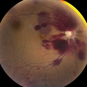
Fundus photograph of a 36-year-old-woman with a preretinal subhyaloid hemorrhage (valsalva retinopathy).
Photographer: Mitzy Torres Soriano
Condition/keywords: macular hemorrhage, premacular hemorrhage, preretinal hemorrhage, subhyaloid hemorrhage, valsalva retinopathy
-
 Subretinal Hemorrhage Due to SRNVM, Fluorescein Angiogram Photograph
Dec 1 2016 by James B. Soque, CRA, OCT-C, COA, FOPS
89-year-old white male with NVAMD and new subretinal hemorrhage, fluorescein angiogram, early phase, of the right eye. Currently receiving anti VEGF treatment.
Photographer: James Soque, CRA, OCT-C, COA, Island Retina, Shirley, NY
Imaging device: Topcon TRC 50 DX, with MERGE software
Condition/keywords: hemorrhage, Hot spot, neovascular age-related macular degeneration (AMD), subretinal hemorrhage, subretinal blood, wet age-related macular degeneration (wet AMD)
-
 SLE Retinopathy
Nov 14 2016 by Mitzy E Torres Soriano, MD
25-year-old female patient with systemic lupus erythematosus. Photographs show cotton wool spots, intraretinal hemorrhages and vascular tortuosity. FA demonstrated retinal vasculitis and OCT revealed cystoid macular edema. In this case diagnosis of SLE was made after ocular manifestation.
Photographer: Grupo Laser Vision, Rosario, Argentina
Condition/keywords: cotton wool spots, occlusive retinal vasculitis, occlusive vasculitis, systemic lupus erythematosus, vasculopathy
-
 Pancytopenia
Mar 24 2016 by Mitzy E Torres Soriano, MD
Fundus photographs of a 35-year-old woman with pancytopenia.
Photographer: Mitzy E. Torres Soriano, Hospital Central de Maracay, Venezuela
Condition/keywords: pancytopenia, retinal hemorrhage
 Loading…