Search results (78 results)
-
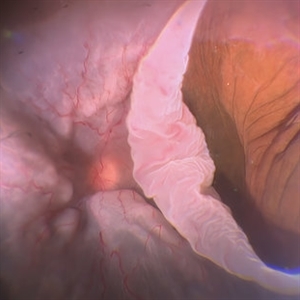
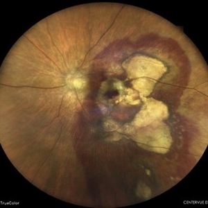
 Hemorrhagic Choroidals
Jan 22 2025 by Danish Shabbir, Ophthalmic Technologist
78 year old female complains of suddenly vision decrease 2 days ago.
Photographer: Danish Shabbir,Retina-EyeCare Centre
Imaging device: Optos California
Condition/keywords: choroidal detachment, Retinal Detachment, retinal detachment with choroidal
-
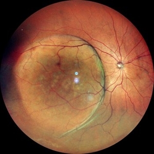
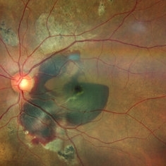
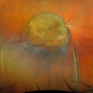 Choroidal Melanoma with Serous Retinal Detachment
Dec 20 2024 by Daniel Davis, OCT-C
67 year old male presenting with large pigmented choroidal mass with serous retinal detachment.
Photographer: Daniel Davis, OCT-C, The Retina Institute
Imaging device: Optos California
Condition/keywords: Retina detachment
-
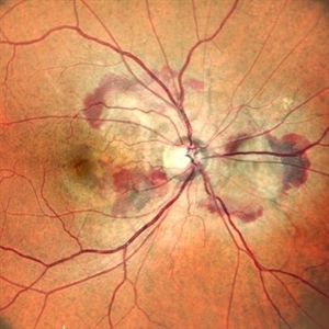 Right Eye Color Photo With Hemorrhages in Case of CNVM With Angioid Streaks
Nov 29 2024 by Anand Temkar
A 45 year old male came with chief complaint of blurring vision in right eyes since past 4 days. His vision is 6/12 in right eye and 6/9 in left eye. His vision was 14 mmHg in right eye and 16 mmHg in left eye. He was diagnosed with Angioid Streaks in both eyes about a year ago, then he developed choroidal neovascularization in his left eye 8 months ago, for which he received AntiVEGF injections x 3. Left eye is a stable eye now. Patient presented with right eye choroidal neovascularization in a case of Angioid Streaks on recent follow up. We have advised him right eye AntiVEGF injections x 3. In this image, the right eye color photo shows bleed from CNVM in case of angioid streaks.
Photographer: Dr.Anand Temkar- Retina Foundation, Ahmedabad
Imaging device: Mirante
Condition/keywords: Angioid Streaks, choroidal neovascular membrane (CNVM)
-
 Choroidal Fracture
Oct 27 2024 by César Adrián Gómez Valdivia, MD
Fundus photograph of a traumatic choroidal fracture & extra-macular sub-retinal hemorrhage.
Photographer: @eyemissu2
Imaging device: TOPCON TRC-50DX
Condition/keywords: Choroidal Fracture
-
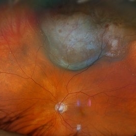
 New Choroidal Melanoma with Exudative Detachment
Oct 16 2024 by Virginia Gebhart
56 year old male with a large pigmented tumor with an exudative detachment inferior and shallow fluid through the macula. Pt states they have been having symptoms for over a year. Scheduled for brachytherapy.
Photographer: Virginia Gebhart, Retina Consultants of Carolina
Imaging device: Optos California
Condition/keywords: Choroidal melanoma, exudative detachment, melanoma
-
 Suprachoroidal Hemorrhage
Dec 3 2024 by Dibya Prabha
Colour Fundus photograph of 62 Year old female patient with Suprachoroidal hemorrhage post trauma
Photographer: Dibya Prabha, LV Prasad eye Institute, Hyderabad
Condition/keywords: suprachoroidal hemorrhage
-
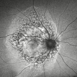
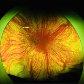 Foveal Hypoplasia / Ocular Albinism
Aug 29 2024 by César Adrián Gómez Valdivia, MD
Fundus photograph of a 64-year-old female patient with foveal hypoplasia, ocular albinism and pendular nystagmus. Findings were bilateral. Retinal and choroidal vasculature are exquisitely beautiful.
Photographer: @eyemissu2
Imaging device: California ICG OPTOS
Condition/keywords: foveal hypoplasia, ocular albinism
-
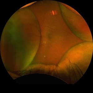
 Idiopathic Uveal Effusion Syndrome
Aug 22 2024 by Jordyn Beckman
61 year old male with Idiopathic Uveal Effusion Syndrome with starry night appearance on fluorescein. 3 weeks s/p single external drainage retinotomy and 9 weeks of oral pred with recurrent choroidal effusions. Has since returned to surgery for secondary drainage retinotomy; subretinal fluid remain persistent.
Photographer: Jordyn Beckman
Imaging device: Optos California
Condition/keywords: chorioretinitis, Choroidal, exudative detachment, window defect
-
 Choroidal Metastasis With Orange Pigment in a Patient With Endometrial Carcinoma
Aug 8 2024 by Guilherme Sturzeneker, MD, MSc
Ultra-widefield fundus photograph and autofluorescence of a 62-year-old woman with endometrial cancer, denoting choroidal metastasis with unusual orange pigment. This presentation is a reminder that the development of orange pigment is not pathognomonic for choroidal melanoma, as it may be seen in other lesions such as carcinoma metastasis.
Photographer: Andrea Almeida
Imaging device: Optos Silverstone
Condition/keywords: choroidal metastasis, metastatic cancer, orange pigment
-
 Choroidal Melanoma
Aug 8 2024 by Virginia Gebhart
88 year old male with new bilobed choroidal melanoma. Pt scheduled for brachytherapy
Photographer: Virginia Gebhart
Imaging device: Optos California
Condition/keywords: melanoma
-
 Choroidal Effusion
Jul 28 2024 by SHILPI H NARNAWARE, ICO ( Retina) , FAICO ( Vitreo-Retina)
Fundus photo of a 53 year old female with 360 degrees choroidal effusion.
Photographer: Shilpi Narnaware, Sarakshi Netralaya , Nagpur, Maharashtra , India
Imaging device: Mirante ( by Nidek)
Condition/keywords: Choroid
-
 Fish Hook Eye Trauma
Jun 12 2024 by Miguel Brito, MD, FASRS
Fundus photograph of a 15-year-old boy post cataract aspiration, pars plana vitrectomy, suprachoroidal drainage, and retinal reattachment surgery secondary to traumatic endophthalmitis.
Photographer: Miguel Brito
Condition/keywords: endophthalmitis, PFCL, Retinal detachment under Silicon Oil, retinal fold
-
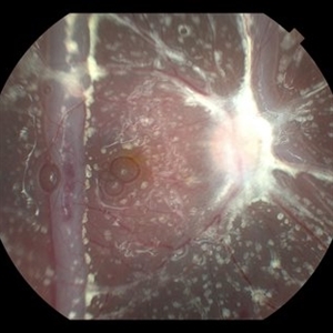
 Giant Retinal Tear with Choroidal Detachment
Jun 12 2024 by Anand Temkar
Intra operative still of a 34 year old male showing Giant Retinal Tear with Choroidal Detachment.
Photographer: Dr.Anand Temkar- Retina Foundation, Ahmedabad
Condition/keywords: choroidal detachment, giant retinal tear
-
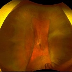
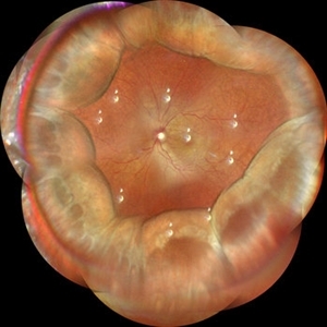
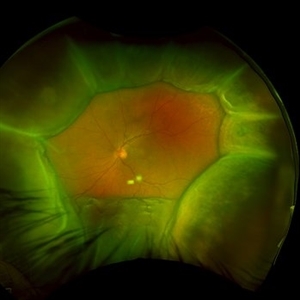
 Uveal Effusion Syndrome
Oct 23 2023 by Gustavo Aguirre-Suarez
Fundus photograph of a 58-year-old man with Type 1 Uveal Effusion Syndrome, showing 360º bullous choroidal detachment.
Photographer: Dr. Gustavo Aguirre-Suarez
Imaging device: Zeiss Clarus 700
Condition/keywords: choroidal effusion, idiopathic uveal effusion syndrome
-
 Choroidal Mass
Sep 21 2023 by Vaidehi Sathaye
Widefield photograph of RE of a 68 year male with choroidal mass.
Photographer: Dr. Vaidehi Sathaye
Imaging device: Mirante
Condition/keywords: choroidal mass
-
 Choroidal Detachment
Aug 14 2023 by Omar Toncel Churio
Fundus photograph of a woman patient with a choroidal detachment.
Photographer: Omar Toncel Churio, Hospital Militar de Especialidades Oftalmológicas, Ciudad de México
Imaging device: Optos California Retinal Camera
Condition/keywords: choroid, detachment, retina
-
 Idiopathic Uveal Effusion Syndrome
Jun 13 2023 by Ahmad B. Tarabishy, MD
66 year old male presented with a 4 month vision of painless decreased vision in the left eye. Clinical findings consistent with idiopathic uveal effusion syndrome. Fundus photography shows 360 degree choroidal elevation with dependent inferior subretinal fluid.
Photographer: Dr. Angela Rico, Retina Specialists of Tampa
Imaging device: Idiopathic Uveal Effusion Syndrome
Condition/keywords: idiopathic uveal effusion syndrome, uveal effusion
-
 Subretinal Hemorrhage
Feb 28 2023 by Akansha Sharma
Color fundus photograph of an 84-year old male with subretinal hemorrhage associated with areas of scarring.
Photographer: Dr. Urmil Shah, Dr. Denish Patel, Dr. Akansha Sharma, Bharati Eye Hospital, Ahmedabad, Gujarat
Condition/keywords: choroidal neovascularization (CNV), subretinal hemorrhage
-
 Oculocutaneous Albinism
Jan 22 2023 by Pietro Dechichi
Fundus photograph of a 6-year-old girl with oculocutaneous albinism. Patient's nystagmus made it difficult to perform the exam. Foveal hypoplasia and evident choroidal vessels can be seen in the retinography
Photographer: Pietro Dechichi
Imaging device: Optos California
Condition/keywords: childhood, foveal hypoplasia, ocular albinism
-
 Choroidal Melanoma with Exudative Retinal Detachment
Mar 2 2023 by Aditya S Kelkar, MS, FRCS, FASRS,FRCOphth
Color fundus photograph of the left eye of a 45 year old male showing choroidal melanoma with exudative retinal detachment.
Photographer: Dr. Pranali Surawase, National Institute of Ophthalmology, Pune, India.
Imaging device: Zeiss Clarus 500
Condition/keywords: choroidal mass, exudative retinal detachment, Retinal detachment
-
 Kissing Serous Choroidal Detachment
Mar 8 2023 by Annaka Gooding
Ultra-widefield fundus photograph of a 73 year old male with a Kissing serous choroidal detachment affecting his right eye. Patient presented at the office following a XEN implant and his vision was sc20/100 PH20/50+1. The physician recommended to start Prednisone treatment.
Photographer: Annaka Gooding
Imaging device: Optos California
Condition/keywords: fundus photography, Kissing Serous Choroidal Detachment, Optos, Right Eye, ultra-wide field imaging
-
 Choroidal Melanoma
Nov 3 2022 by pedro fernandes souza neto
Transillumination of Enucleation specimen of Choroidal Melanoma: anterior chamber is closed. Total secondary retinal detachment with subretinal serous fluid and some subretinal hemorrhages are present.
Photographer: Eduardo Marback, Federal University of Bahia, Brazil
Condition/keywords: enucleation, melanoma
-
 Submacular Hemorrhage PCV
May 6 2022 by Shobhit Chawla, M.S.
Submacular hemorrhage in a 38 years old female patient cause polyp bleed in PCV.
Photographer: Shobhit Chawla
Imaging device: Zeiss Clarus 500
Condition/keywords: polypoidal choroidal vasculopathy (PCV), submacular hemorrhage
-
 Angioid Streaks
Jun 14 2022 by Kingston Rodolfo Ureña-Wong, MD, Opht, MSc
Fundus photograph of an 26-year-old woman with pseudoxanthoma elasticum and angioid streaks. She developed a choroidal neovascular membrane which was treated with anti-VEGF successfully.
Photographer: Kingston Rodolfo Ureña-Wong, Asociación para evitar la ceguera en México, México.
Imaging device: Zeiss Clarus
Condition/keywords: Angioid Streaks
-
 Perforating Ocular Trauma and Choroidal Rupture due to Shotgun Pellet
Mar 31 2022 by Franco Benvenuto, MD
Fundus photograph of a 17-year-old with shotgun injuries with numerous metal pellets in the chest, neck, brain, and face. Fundus exploration showed the left globe with posterior-inferior eye rupture, vitreous hemorrhages and choroidal rupture.
Photographer: Franco Benvenuto, Universidad de Buenos Aires, Argentina. Universidad de Guadalajara, México.
Condition/keywords: choroidal rupture, penetrating trauma, shotgun
 Loading…