Search results (2839 results)
-
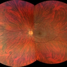
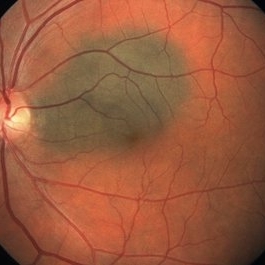
 Siegrist Streaks
Mar 29 2013 by Henry J. Kaplan, MD
Typical Siegrist streaks in hypertensive choridopathy; hyperpigmentations in a linear fashion along choroidal vessels , a rare finding.
Condition/keywords: hypertensive choroidopathy, Siegrist Streaks
-
---thumb.jpg/image-square;max$300,300.ImageHandler) Myopic fundus
Myopic fundus
Jan 11 2013 by Hyung-Woo Kwak, MD
Myopic fundus reveals yellow-colored lacquer cracks and peripapillary atrophy. There was visible choroidal vessel due to thin retina.
Photographer: Misook Lee, Kyung Hee Univsersity Hospital, Seoul
Imaging device: Zeiss f 450 plus
Condition/keywords: myopic fundus
-
 Tigroid Fundus
Aug 31 2021 by Ricardo Leitão Guerra
True color (RGB) confocal scanning laser ophthalmoscopy of a 37-year-old male with myopia highlighting choroidal vessels and vortex veins.
Photographer: Juliana Rio, MD. Leitão Guerra - Oftlamologia, Salvador - Brazil
Imaging device: Zeiss Clarus 7000
Condition/keywords: myopia, retina, tigroid fundus, vortex vein
-
 Vortex Vein In A Patient With A Blond Fundus
Oct 2 2013 by Jerald A. Bovino, MD
The vortex vein and vortex vein ampulla are visible in this patient with a blonde (lightly pigmented) fundus.
Condition/keywords: choroidal circulation, fundus photograph, vortex vein
-
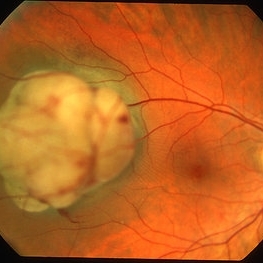
 Macular Choroidal Osteoma
Aug 17 2012 by Jonathan L. Prenner, MD
Macular choroidal osteoma in a 29-year-old woman
Condition/keywords: macular choroidal osteoma
-
 Geographic Atrophy, Fundus photograph
Aug 23 2012 by Gerardo Garcia-Aguirre, MD
Fundus photograph of an 85-year-old patient with age related macular degeneration and geographic atrophy. A large area with well-defined borders is observed, in which the choroidal vasculature is visualized.
Photographer: Noemí Hernández, Asociación para Evitar la Ceguera en México
Imaging device: Zeiss FF4
Condition/keywords: geographic atrophy
-
 Choroidal melanoma case 4 - partly amelanotic
Jan 11 2013 by Alex P. Hunyor, MD
Choroidal melanoma with amelanotic "collar stud."
Condition/keywords: melanoma
-
 Choroidal Melanoma
Jul 4 2012 by John T. Thompson, MD
Amelanotic choroidal melanoma with serous retinal detachment
Condition/keywords: choroidal tumor, exudative retinal detachment, melanoma
-
 Myopic CNV
Jan 11 2013 by Alex P. Hunyor, MD
Myopic macular degeneration complicated by subretinal neovascularisation, left eye.
Condition/keywords: high myopia, myopia, myopic choroidal neovascularization (CNV)
-
 Myopic Choroidal Neovascular Membrane
Mar 25 2013 by Ratimir Lazic, MD, PhD
Color fundus photography of a 33-year-old female. In macular area subretinal hemorrhage can be seen. Area of atrophy temporal from PNO. Myopic changes of posterior pole and mid periphery can be noticed. The patient has been treated with 2 consecutive ranibizumab intravitreal injections. BCVA at baseline was 0,05 (Snellen lines) and 0,3 (Snellen lines) 2 months after.
Photographer: Marko Lukic, MD
Imaging device: Zeis Visucam Lite 2
Condition/keywords: high myopia, myopic choroidal neovascularization (CNV), ranibizumab
-
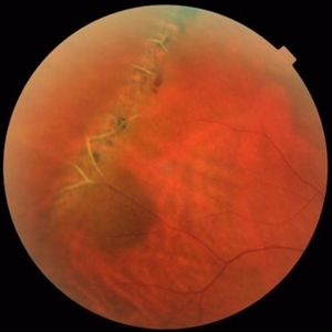 Lattice Degeneration and Choroidal Nevus
Oct 10 2015 by Hamid Ahmadieh, MD
Color fundus photograph of the right eye of a 46-year-old woman with a typical lattice degeneration and an adjacent choroidal nevus.
Photographer: Solmaz Shahmohammad, Negah Eye Center, Tehran, Iran
Condition/keywords: choroidal nevus, color fundus photograph, lattice degeneration
-
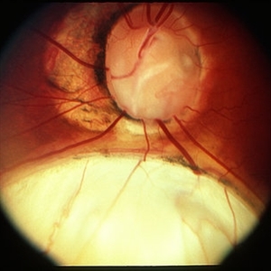 Coloboma
Mar 29 2013 by Henry J. Kaplan, MD
Optic disc and inferonasal choroidal coloboma in the same patient #2.
Condition/keywords: coloboma, coloboma of choroid, coloboma of optic disc
-
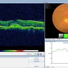
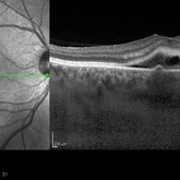
 Polypoidal Choroidal Vasculopathy-OCT
Aug 27 2012 by Young Hee Yoon, MD, PhD
SD-OCT image of a 56-year-old woman. Her best-corrected visual acuity was 20/30.
Photographer: Kyoung Ree Kim, Asan Medical Center
Imaging device: Heidelberg Spectralis
Condition/keywords: polypoidal choroidal vasculopathy (PCV)
-
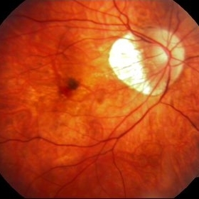
 Choroidal melanoma case 3 - peripapillary
Jan 11 2013 by Alex P. Hunyor, MD
Right peripapillary choroidal melanoma.
-
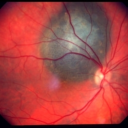
 Melanocytoma with Choroidal Melanoma
Oct 8 2012 by Susanna S. Park, MD, PhD
Fundus photograph of a 75-year-old woman with a slowly growing pigmented lesion.
Photographer: Ellen Redenbo, University of California Davis Eye Center
Condition/keywords: melanocytoma
-
 Choroidal Osteoma Plus CNV
Sep 2 2012 by Hamid Ahmadieh, MD
Color fundus photograph and OCT imaging of a 47-year-old man with a juxtafoveal CNV superimposed on a choroidal osteoma.
Photographer: Hamid Ahmadieh, Ophthalmic Research Center, Labbafinejad Medical Center
Imaging device: Topcon
Condition/keywords: choroidal neovascularization (CNV), choroidal osteoma, optical coherence tomography (OCT)
-
 Diffuse Choroidal Melanoma OCT
Aug 24 2012 by John S. King, MD
Photographer: Kristin Konecki, OcuSight Eye Care Center, Rochester, NY
-
 Punctate Inner Choroidopathy with CNV Treated with Bevacizumab # 6 of 7
Feb 28 2013 by Gregory R. Blaha, MD, PhD
Fundus photo following treatment with bevacizumab in a 31-year-old female with vision loss from a choroidal neovascular membrane (CNV) from punctate inner choroidopathy. The vision improved and was stable following a single injection.
Photographer: Gerard Gauthier, Spindel Eye Assoc., Derry, NH
Imaging device: Zeiss FF 450 Plus
Condition/keywords: bevacizumab, choroidal neovascularization (CNV), punctate inner choroidopathy (PIC)
-
 Sturge-Weber Episcleral-Vessels
Apr 17 2014 by Susanna S. Park, MD, PhD
External photo of the right eye of this 8-year-old Hispanic boy with Sturge -Weber Syndrome and diffuse choroidal hemangioma showing dilated episcleral vessels.
Photographer: Ellen Redenbo, University of California Davis Eye Center
Condition/keywords: dilated episcleral vessels, Sturge-Weber syndrome
-
 Choroidal Nevus
May 8 2014 by S. Natarajan, MD, FASRS, FRCS (GLASGOW) , FICO, D.Sc, FELA
Fundus photograph of a 48-year-old female in for a routine eye checkup with vision 6/6 OU. Showed solitary pigmented juxtapapilary nevus of 3*2 disc diameters size.
Photographer: ADITYA JYOT EYE HOSPITAL,MUMBAI INDIA
Condition/keywords: choroidal nevus
-
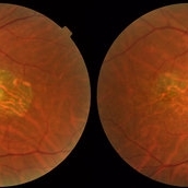 Central Areolar Choroidal Dystrophy
Jul 7 2015 by Hamid Ahmadieh, MD
Color fundus photograph of both eyes of a 58-year-old man with progressive loss of vision. VA OD is 20/60 and VA OS is 20/400.
Photographer: Soulmaz Shahmohammad, Negah Eye Center, Tehran, Iran
Imaging device: Topcon
Condition/keywords: central areolar choroidal dystrophy (CACD), color fundus photograph
-
 Choroidal Detachment, In Stereo
Sep 25 2012 by Michael P. Kelly, FOPS
Photographer: Michael P. Kelly, FOPS Director, Duke Eye Labs, Duke University Hospital, Duke Eye Center, Durham, NC
Imaging device: Zeiss FF3C
Condition/keywords: choroidal detachment, stereo pair
-
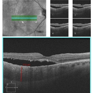
 Recurrent Central Serous Choroidopathy
Aug 21 2012 by Edwin H. Ryan, MD
EDI-OCT showing thickened choroid and subretinal fluid
Photographer: Edwin Ryan Jr. MD, VitreoRetinal Surgery, PA
Imaging device: Heidelberg Spectralis
Condition/keywords: central serous chorioretinopathy (CSCR), choroidal thickening, enhanced depth imaging
-
 Choroidal metastasis - case 4
Jan 11 2013 by Alex P. Hunyor, MD
Choroidal metastasis from breast carcinoma, with exudative detachment of the macula.
Condition/keywords: choroidal metastasis
-
 Pigment Epithelial Detachment late FA with small occult CNV
Jul 6 2012 by Tarek S. Hassan, MD, FASRS
72-year-old man with VA loss and metamorphopsia of 2 months duration. PED found, testing done to rule out CNV. Very suspicious for CNV in superonasal fovea/parafovea.
Condition/keywords: choroidal neovascularization (CNV), pigment epithelial detachment (PED)
 Loading…