Search results (2839 results)
-
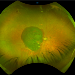
 Tuberculoma
Jun 4 2025 by Paulina Araujo
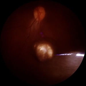
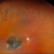
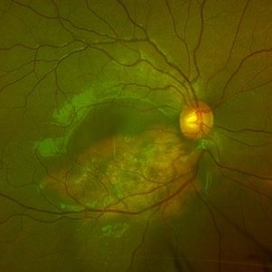
The 55-degree central fundus photograph of the left eye reveals an elevated, nodular whitish choroidal lesion along the inferior temporal arcade, consistent with a tuberculoma.
Photographer: Paulina D.Araujo Martínez, Asociación para Evitar la Ceguera en México I.A.P., Hospital Dr Luis Sánchez Bulnes.
Condition/keywords: Choroidal-tuberculoma
-
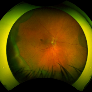
 Choroidal Nevus
Jun 4 2025 by Paulina Araujo
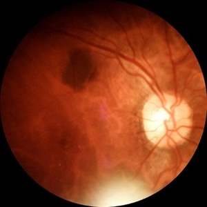
The 55-degree central fundus photograph of the right eye reveals a choroidal nevus measuring 0.5 disc diameters along the superior temporal arcade.
Photographer: Paulina D.Araujo Martínez, Asociación para Evitar la Ceguera en México I.A.P., Hospital Dr Luis Sánchez Bulnes.
Condition/keywords: Choroidal nevus
-
 Choroidal Rupture
Jun 4 2025 by Paulina Araujo
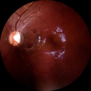
The 55-degree central fundus photograph of the left eye reveals a choroidal rupture in the nasal parafoveal area secondary to blunt ocular trauma.
Photographer: Paulina D.Araujo Martínez, Asociación para Evitar la Ceguera en México I.A.P., Hospital Dr Luis Sánchez Bulnes.
Condition/keywords: choroidal rupture
-
 Multi-modal Imaging of Type - 1 CNVM
May 30 2025 by Shivankar Sen
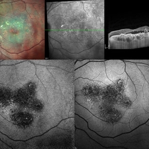
Multimodal Imaging of a case of Polypoidal Choroidal Vasculopathy Multicolor Reflectance showing multiple green-hyper-fringent lesions in the macular region (Up Left) Infra-red Autofluorescence and Blue Autofluorescence showing hypo-autofluorescent areas correspondingly revealing the exact extent of the sub-RPE Lesion (Down left and right respectively) Optical Coherence Tomography - Enhanced Depth Imaging showing Thumb-shaped Pigment Epithelial Detachment with presence of Sub-retinal fluid and Hyper-reflective foci (Top Right)
Photographer: Dr. Shivankar Sen
Imaging device: Heidelberg Spectralis HRA+OCT
Condition/keywords: Blue autofluroscence, CNVM, multicolor, near infrared autofluorescence (NIRAF), PCV, reflectance
-
 Radiation Retinopathy with BRVO
May 28 2025 by Virginia Gebhart
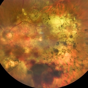
46 year old male with regressing choroidal melanoma. Stable pigment dispersion over biopsy site, BRVO secondary to radiation retinopathy. BCVA CF, will continue to observe.
Photographer: Virginia Gebhart, Retina Consultants of Carolina
Imaging device: Optos California
Condition/keywords: brachytherapy, branch retinal vein occlusion (BRVO), BRVO, Choroidal melanoma, melanoma, radiation retinopathy
-
 Macular Star
May 27 2025 by César Adrián Gómez Valdivia, MD
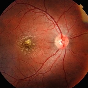
Macular Star found in a 31 year-old male patient with suspected Cat Scratch Disease. Typical intraocular presentations include neuroretinitis with optic nerve edema, macular star formation, and discrete white retinal or choroidal lesions. Findings were unilateral.
Photographer: @eyemissu2
Imaging device: TOPCON TRC-50DX
Condition/keywords: macular star
-
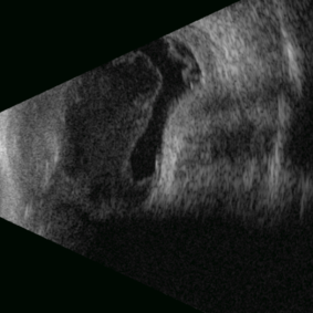
 Glass
Glass
May 21 2025 by Gustavo Uriel Fonseca Aguirre
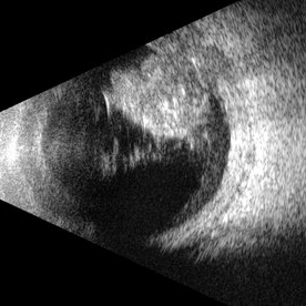
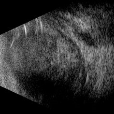
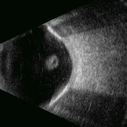
This B-mode transverse ultrasound scan reveals an intraocular glass foreign body secondary to penetrating trauma, with associated vitreous and subhyaloid hemorrhage. The glass fragments appear as hyperechoic linear structures in both the vitreous cavity and the retinachoroidal complex.
Photographer: Gustavo U. Fonseca Aguirre, Hospital Conde de Valenciana, Ciudad de México
Condition/keywords: glass, intraocular foreign body
-
 PCV
PCV
May 20 2025 by LUBNA AHMAD
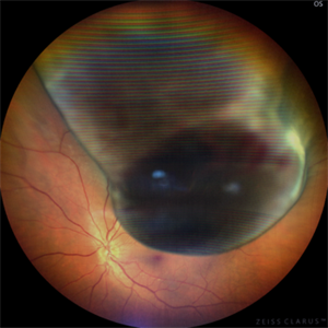
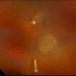
Fundus image of a 66 year old male with huge branching vascular network, fresh hemorrhage and scarred PCV lesion.
Photographer: Sana Zamani
Imaging device: zeiss clarus 500
Condition/keywords: branching vascular network (BVN), polypoidal choroidal vasculopathy (PCV)
-
 Scleral Rupture
May 9 2025 by Gustavo Uriel Fonseca Aguirre
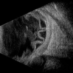
This B-mode longitudinal ultrasound scan reveals dense vitreous hemorrhage, subretinal fluid, annular choroidal detachment, and scleral wall discontinuity with adjacent scleral folds. These findings indicate severe ocular trauma with probable scleral rupture and multi-compartment involvement.
Photographer: Gustavo U. Fonseca Aguirre, Hospital Conde de Valenciana, Ciudad de México
Condition/keywords: ocular trauma, scleral rupture
-
 Traumatic Retinal Detachment
May 5 2025 by Gustavo Uriel Fonseca Aguirre
This B-mode longitudinal ultrasound scan over the macular area reveals vitreous hemorrhage, retinal detachment with folding, peripheral annular choroidal detachment, and sub-Tenon's fluid in the setting of blunt ocular trauma. The findings indicate severe posterior segment disruption with multi-compartment involvement.
Photographer: Gustavo U. Fonseca Aguirre, Hospital Conde de Valenciana, Ciudad de México
Condition/keywords: blunt trauma, Retinal Detachment
-
 Uveal Melanoma
Apr 26 2025 by Vishal Agrawal, MD, FRCS,FACS,FASRS
A 32 year-old male presented with complaints of perceiving a shadow in OS for 15-20 days. His BCVA was 20/20 OU. On Fundus examination, a large, elevated, well-defined, pigmented choroidal mass with few hemorrhages over the lesion was seen and a provisional diagnosis of uveal melanoma was made. urgent oncological consultation was recommended for further treatment.
Photographer: Dr Ayushi Gupta
Imaging device: Clarus 700
Condition/keywords: melanoma
-
 Sclerochoroidal Calcification
Apr 24 2025 by Virginia Gebhart
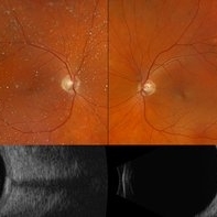
70 year old male referred for amelanotic lesion in the STA OU. Ultrasound shows slightly elevated lesions with hyperreflectivity and posterior shadowing with reduplication artifact consistent with sclerochoroidal calcification. Recommend yearly observation.
Photographer: Virginia Gebhart, Retina Consultants of Carolina
Imaging device: Optos California, Ellex Eye Cubed
Condition/keywords: asteroid hyalosis, B scan ultrasound, sclerochoroidal calcification
-
 Posterior Nodular Scleritis
Apr 23 2025 by Gustavo Uriel Fonseca Aguirre
This B-mode ultrasound scan demonstrates a posterior scleral nodule accompanied by vitritis, serous retinal detachment, and annular choroidal detachment. The nodule appears as a localized hypoechoic scleral thickening, while the serous retinal detachment shows a smooth convex configuration. The choroidal detachment presents with the characteristic ring-shaped elevation, suggesting significant intraocular inflammation or hypotony.
Photographer: Gustavo U. Fonseca Aguirre, Hospital Conde de Valenciana, Ciudad de México
Condition/keywords: posterior nodular scleritis, posterior scleritis
-
 Sub retinal Bleed
Apr 23 2025 by Anjana Mirajkar, MS Ophthalmology
A widefield image of right eye of a 65 year old male showing large subretinal bleed at the posterior pole most likely in a case of PCV.
Photographer: Dr. Anjana Mirajkar- HV desai eye hospital ,Pune
Imaging device: optos
Condition/keywords: idiopathic polypoidal choroidal vasculopathy, subretinal blood
-
 Serous Choroidal Detachment
Apr 18 2025 by Tracy Vu
Fundus photograph of a 77-year-old male with near 360 degree choroidal effusions accompanied by a mild serous choroidal detachment in the inferotemporal quadrant.
Condition/keywords: choroidal effusion, serous choroidal detachment, serous retinal detachment
-
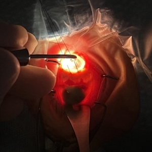 Intraoperative Transillumination of Choroidal Melanoma
Apr 18 2025 by Virginia Gebhart
Intraoperative photo of transillumination of choroidal melanoma before plaque placement in 36 year old female.
Photographer: Chris Bergstrom, MD, OD
Imaging device: iphone
Condition/keywords: choroidal melanoma, intraoperative, transillumination
-
 Choroidal Osteoma
Apr 17 2025 by Gustavo Uriel Fonseca Aguirre
Scanning laser ophthalmoscopy reveals a well-circumscribed, yellowish-white choroidal osteoma overlying the macular region and extending into the inferior temporal vascular arcade. Retinal vessels course normally over the tumor surface, with no evidence of subretinal fluid or hemorrhage. The surrounding retina shows preserved architecture without secondary degenerative changes.
Photographer: Gustavo U. Fonseca Aguirre, Hospital Conde de Valenciana, Ciudad de México
Condition/keywords: choroidal osteoma, macular choroidal osteoma
-
 Choroidal Osteoma
Apr 17 2025 by Gustavo Uriel Fonseca Aguirre
Top (B-mode): The longitudinal scan reveals a hyperechoic, flat, and well-demarcated macular lesion with posterior acoustic shadowing, pathognomonic for choroidal osteoma. Bottom (A-mode): Standardized tracing shows a tall initial spike (100% reflectivity) at the tumor surface with rapid decay to acoustic silence, confirming sound absorption by calcified tissue. This pattern remains unchanged at variable gain settings.
Photographer: Gustavo U. Fonseca Aguirre, Hospital Conde de Valenciana, Ciudad de México
Condition/keywords: choroidal osteoma, macular choroidal osteoma
-
Pseudomelanoma (PEHCR)
Apr 15 2025 by Virginia Gebhart
67 year old male referred for peripheral choroidal lesion. Clinical exam and Bscan findings consistent with a subRPE hemorrhage secondary to peripheral exudative hemorrhagic chorioretinopathy. No vascularity on ultrasound. OD has small subRPE hemorrhage as well. Pt is on Eliquis. Will monitor with serial exams. Sponsored by the number 2
Photographer: Virginia Gebhart, Retina Consultants of Carolina
Imaging device: Optos California
Condition/keywords: peripheral exudative hemorrhagic chorioretinopathy (PEHCR)
-
Hemorrhagic Choroidal Detachment
Apr 14 2025 by Gustavo Uriel Fonseca Aguirre
This B-mode transverse ultrasound scan demonstrates a hemorrhagic choroidal detachment with a characteristic wreath-like configuration, accompanied by concurrent retinal detachment. The choroidal lesion shows dome-shaped elevation with heterogeneous internal reflectivity, while the detached retina appears as a hyperechoic, undulating membrane.
Photographer: Gustavo U. Fonseca Aguirre, Hospital Conde de Valenciana, Ciudad de México
Condition/keywords: hemorrhagic choroidal detachment
-
Ciliary Body Detachment in Uveal Effusion Syndrome
Apr 11 2025 by Siri Uppuluri
Ultrasound biomicroscopy of a phakic left eye in an 82-year-old man demonstrating ciliary body detachment in the setting of uveal effusion syndrome. Patient also presented with 360 choroidal effusions and underwent sclerectomy and drainage of choroidal effusions with resolution after surgical intervention.
Photographer: Siri Uppuluri, MD; Rutgers New Jersey Medical School
Condition/keywords: uveal effusion syndrome
-
360 Choroidal Detachment in Uveal Effusion Syndrome
Apr 11 2025 by Siri Uppuluri
Fundus photograph of a phakic left eye in an 82-year-old man demonstrating 360 choroidal detachment secondary to uveal effusion syndrome. He underwent sclerectomy and drainage of the choroidal effusions with resolution after surgical intervention.
Photographer: Siri Uppuluri, MD; Rutgers New Jersey Medical School
Condition/keywords: choroidal detachment
-
 Open Funnel (Transversal)
Apr 10 2025 by Gustavo Uriel Fonseca Aguirre
This B-mode transverse ultrasound scan reveals a chronic rhegmatogenous retinal detachment, demonstrating a funnel-shaped configuration with a narrow intraluminal space. Two hyperechoic choroidal calcifications are present, indicative of chronicity.
Photographer: Gustavo U. Fonseca Aguirre, Hospital Conde de Valenciana, Ciudad de México
Condition/keywords: open funnel RD, Retina detachment
-
 Treated Melanoma with Iluvien Implant
Apr 9 2025 by Virginia Gebhart
62 year old female 4 mo s/p brachytherapy for amelanotic choroidal melanoma. Iluvien implant given 4 wks s/p plaque removal, lesion is stable with resolved exudative detachment and subretinal fluid
Photographer: Virginia Gebhart, Retina Consultants of Carolina
Imaging device: Optos California
Condition/keywords: amelanotic melanoma, brachytherapy, choroidal melanoma, Iluvien, melanoma
-
 Choroidal Melanoma with Exudative Detachment
Apr 7 2025 by Virginia Gebhart
Autofluorescence image of 36 year old female showing demarcation line of fluid/detachment from new choroidal melanoma. Pt will be scheduled for brachytherapy pending CT scan results.
Photographer: Virginia Gebhart, Retina Consultants of Carolina
Imaging device: Optos California
Condition/keywords: Autoflourescence, autofluorescence imaging, choroidal melanoma, melanoma, retinal detachment
 Loading…