Search results (7 results)
-
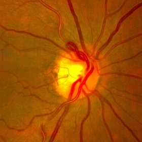 Prepapillary Vascular Loop
Jul 4 2025 by KANWALJEET HARJOT MADAN, M.S. (Ophthalmology); FAICO (Vitreous - Retina)
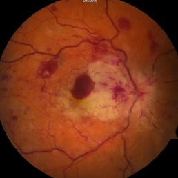
This is the fundus picture of right eye of a young 32 years female depicting pre papillary vascular loop. A prepapillary vascular loop is a congenital anomaly of the optic disc that presents as an elevated and twisted bundle of vessels projecting into the vitreous cavity. It is a benign condition, usually unilateral but can be bilateral. It is asymptomatic and discovered during routine eye examination. This anomaly can sometimes cause complications like branch retinal artery occlusion, vitreous hemorrhage, or sub retinal hemorrhage.
Photographer: Dr. Kanwaljeet Harjot Madan, Thind Eye Hospital, Jalandhar City (Punjab) INDIA.
Imaging device: Zeiss Fundus Camera
Condition/keywords: branch retinal artery occlusion (BRAO), optic disc, Prepapillary Vascular Loop, SUB RETINAL HEMORRHAGE, Vitreous hemorrhage
-
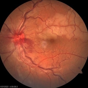 Combined central retinal vein occlusion and branch retinal arteriolar occlusion
Sep 13 2022 by Ruchir Mehta, DO, DNB, FRCS
Fundus photograph of left eye of a 63 years old female with known type 2 DM and HTN showing combined central retinal venous occlusion and superior branch retinal arteriolar occlusion
Photographer: Ruchir Mehta, Mehta Superspeciality Eye Hospital, Jamnagar, Gujarat, India
Imaging device: Zeiss Visucam 500
Condition/keywords: branch retinal artery occlusion (BRAO), central retinal vein occlusion (CRVO), COMBINED
-
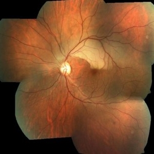
 Laser Induced BRAO in IRVAN Syndrome
May 3 2019 by Deependra Vikram Singh, MD FASRS
Fundus photograph of a 26-year-old man with IRVAN syndrome referred for vitreous surgery in OS for secondary rhegmatogenous retinal detachment. OD has received laser photocoagulation for capillary nonperfusion areas and retinal artery macroaneurysm associated with retinal vasculitis. Fundus photograph of OD shows laser induced nasal BRAO. Case re-emphasizes why laser for macroaneurysm should be avoided in cases with IRVAN.
Photographer: Deependra V Singh, Eye-Q Superspecialty Eye Hospitals. Gurugram, India
Imaging device: Zeiss Visucam 500
Condition/keywords: arteriolar macroaneurysm, branch retinal artery occlusion (BRAO), laser photocoagulation
-
 Branch Retinal Artery Occlusion With Calcium Embolus at the Disc - Fundus Photo
Apr 7 2018 by Rameez N Hussain, MD
Acute retinal artery occlusion with a calcium embolus at the disc and retinal whitening.
Photographer: DR RAMEEZ N HUSSAIN
Imaging device: zeiss
Condition/keywords: branch retinal artery occlusion (BRAO), embolus, fundus photograph, retinal edema
-
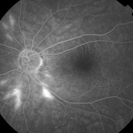 Susac's Syndrome
Feb 13 2018 by John S. King, MD
Background: 46-year-old WF with CML (stable on Sprycel) saw her PCP for headaches without known cause; Headaches worsened and became confused, disoriented, off balance, and impaired short term memory. Heme-oncology ordered MRI that showed abnormal signal in the cerebellum and other parts of the brain, and LP has elevated protein. LP did show positive tau test, but fortunately, was a false positive for CJD. IV and PO steroids started and symptoms improved. MRI showed much improvement one month since starting steroids. 3 weeks later had a scotoma in right eye and eye doctor did not find anything at that time to cause it. Tinnitus developed (and some intermittent vertigo before that) and ENT referred back to eye doctor, who then referred the patient to Dr. Zocchi. He found a CWS and BRAO OD, and bilateral arteritis. She had some additional work-up for vasculitis. Given the retinal arteritis, cochlear issues, and MRI findings, Dr.Zocchi suspected Susac's Syndrome. She was started on multiple regimens including prednisone, IVIG, azathiprine, and MTX, and has had the best reponse to IVIG (FA shows a recurrence/worsening while adjusting IMT). She is stable and doing well with 20/20 vision in both eyes.
Photographer: Kay Dalby
Imaging device: Topcon
Condition/keywords: retinal vasculitis, Susac's syndrome
-
 Branch Retinal Artery Occlusion With Concurrent Central Retinal Vein Occlusion
Oct 5 2016 by Larry M Puthenparambil, MD
Branch retinal artery occlusion with concurrent central retinal vein occlusion.
Photographer: Stacey Groom
Imaging device: Topcon
Condition/keywords: branch retinal artery occlusion (BRAO), central retinal vein occlusion (CRVO)
-
 BRAO
BRAO
Jun 29 2014 by John S. King, MD
BRAO.
Photographer: Wayne A Ladlee Jr
Condition/keywords: branch retinal artery occlusion (BRAO), embolic
 Loading…