Search results (185 results)
-
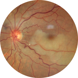
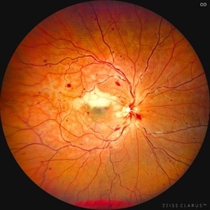
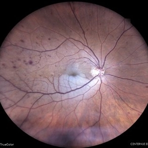
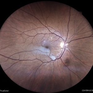
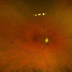
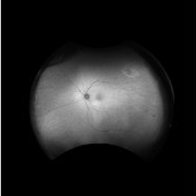
 Branch Retinal Artery Occlusion
Dec 19 2025 by Gayathri M S
Multicolor Reflectance and Blue Reflectance of a 55 yr male patient with blurring since 1 month shows classical sectoral retinal whitening.
Photographer: Gayathri MS
Imaging device: Heidelberg Spectralis
Condition/keywords: blue reflectance, branch retinal artery occlusion (BRAO), multicolor
-
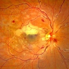
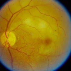
 Hollenhorst Plaque
Sep 2 2025 by KANWALJEET HARJOT MADAN, M.S. (Ophthalmology); FAICO (Vitreous - Retina)
A 64 year-old male presented with sudden decrease in vision in LE for 1 week. His BCVA in LE was 20/200. Fundus exam revealed presence of whitish ischemic area in macula superior to fovea suggestive of branch retinal artery occlusion. A bright tiny refractile cholesterol embolus (Hollenhorst plaque) was visible in retinal artery. The patient was advised cardiology consultation.
Photographer: Dr. Kanwaljeet Harjot Madan, Thind Eye Hospital, Jalandhar City (Punjab). INDIA.
Imaging device: Zeiss Fundus Camera
Condition/keywords: branch retinal artery occlusion (BRAO), hollenhorst plaque
-
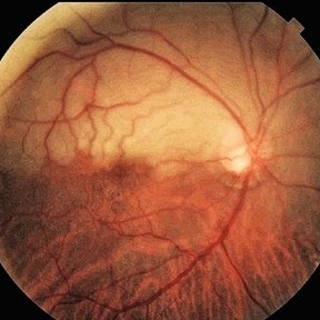
 Prepapillary Vascular Loop
Jul 4 2025 by KANWALJEET HARJOT MADAN, M.S. (Ophthalmology); FAICO (Vitreous - Retina)
This is the fundus picture of right eye of a young 32 years female depicting pre papillary vascular loop. A prepapillary vascular loop is a congenital anomaly of the optic disc that presents as an elevated and twisted bundle of vessels projecting into the vitreous cavity. It is a benign condition, usually unilateral but can be bilateral. It is asymptomatic and discovered during routine eye examination. This anomaly can sometimes cause complications like branch retinal artery occlusion, vitreous hemorrhage, or sub retinal hemorrhage.
Photographer: Dr. Kanwaljeet Harjot Madan, Thind Eye Hospital, Jalandhar City (Punjab) INDIA.
Imaging device: Zeiss Fundus Camera
Condition/keywords: branch retinal artery occlusion (BRAO), optic disc, Prepapillary Vascular Loop, SUB RETINAL HEMORRHAGE, Vitreous hemorrhage
-
 Branch Retinal Artery Occlusion
Oct 1 2024 by Angel Enrique Flores Pineda
Fundus photograph of a 78-year-old woman with poorly controlled systemic arterial hypertension and dyslipidemia. Hollenhorst plaque can be observed.
Photographer: Angel Enrique Flores Pineda, Hospital General de Zona #20
Imaging device: Smartphone (IPhone 15 plus)
Condition/keywords: branch retinal artery occlusion (BRAO)
-
 Hollenhorst Plaque
Jun 25 2024 by Virginia Gebhart
75 year female with complaint of shadow in the bottom of her vision for many years. Hollenhorst plaque on superior pole of the disc and sclerotic superotemporal arteriole. Also DBHs superiorly most likely due to combined BRAO/BRVO.
Photographer: Virginia Gebhart
Imaging device: Topcon 50DX
Condition/keywords: branch retinal artery occlusion (BRAO), branch retinal vein occlusion (BRVO), hollenhorst plaque, sclerotic arteriole
-
 Combined Central Retinal Vein Occlusion with Branch Retinal Artery Occlusion
Apr 29 2024 by KANWALJEET HARJOT MADAN, M.S. (Ophthalmology); FAICO (Vitreous - Retina)
This is fundus photograph of a 46-year male patient who presented with sudden diminution of vision in his right eye (RE) for 3 days. He was hypertensive but non diabetic. On examination, his best corrected vision in RE was 6/12. His left eye (LE) was normal. His fundus examination in RE revealed multiple intra retinal hemorrhages in all quadrants with tortuosity of veins suggestive of central retinal vein occlusion (CRVO) with mild disc edema. An ischemic area was seen superior to fovea suggestive of branch retinal artery occlusion. OCT depicted thickening of inner retinal layers with little evidence of macular edema. Hematological and cardio vascular investigations were done. He had bilateral thickening of intimal and medial walls of carotid arteries. He was under cardiology treatment. His vision improved to 6/6.
Photographer: Dr. Kanwaljeet Harjot Madan, M.S. (Ophthalmologist) Fellow in Vitrous & Retina. Thind Eye Hospital, Jalandhar City. Punjab. India
Condition/keywords: branch retinal artery occlusion (BRAO), central retinal vein occlusion (CRVO)
-
 Combined Central Retinal Vein Occlusion with Branch Retinal Artery Occlusion
Apr 28 2024 by KANWALJEET HARJOT MADAN, M.S. (Ophthalmology) Fellow in Vitreous & Retina
This is fundus photograph of a 46-year male patient who presented with sudden diminution of vision in his right eye (RE) for 3 days. He was hypertensive but non diabetic. On examination, his best corrected vision in RE was 6/12. His left eye (LE) was normal. His fundus examination in RE revealed multiple intra retinal hemorrhages in all quadrants with tortuosity of veins suggestive of central retinal vein occlusion (CRVO) with mild disc edema. An ischemic area was seen superior to fovea suggestive of branch retinal artery occlusion. OCT depicted thickening of inner retinal layers with little evidence of macular edema. Hematological and cardio vascular investigations were done. He had bilateral thickening of intimal and medial walls of carotid arteries. He was under cardiology treatment. His vision improved to 6/6.
Photographer: Dr Kanwaljeet Harjot Madan
Condition/keywords: branch retinal artery occlusion (BRAO), central retinal vein occlusion
-
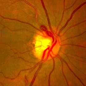
 Superior Hemi-Central Retinal Artery Occlusion
Apr 24 2024 by Mosab Salah
Fundus photograph -inverted view- taken by smartphone fundus photography, of a young man with sudden onset altitudinal field defect, a Superior Hemi-Central Retinal Artery Occlusion noted.
Photographer: Dr Mosab Salah, The Islamic Hospital, Amman, Jordan
Imaging device: smartphone fundus photography and 30 D Lens
Condition/keywords: arterial occlusion, branch retinal artery occlusion (BRAO), BRAO, CRAO, Hemi-Central Retinal Artery Occlusion (CRAO), occlusive vasculitis, smartphone fundus photography
-
 Proliferative Diabetic Retinopathy
Mar 28 2024 by Houda Brarou
Mosaic fundus photograph of the right eye of a 43 years old male patient with a proliferative diabetic retinopathy
Photographer: Houda Braou , Mohammed V military hospital of Rabat
Imaging device: TOPCON DRI OCT Triton Plus
Condition/keywords: diabetes, diabetic retinopathy, proliferative diabetic retinopathy (PDR)
-
 Familial Dominant Drusen
Mar 28 2024 by Houda Brarou
Familial Dominant Drusen is a genetically inherited retinal dystrophy and thought to represent an early-onset variant of age related macular degeneration. The gene responsible is EFEMP1 and inherited in autosomal dominant manner with variable expressivity. It is represented with multiple radially elongated small drusen in early stages and in later stages they become larger and more confluent. Geographic atrophy occurs in advanced stages.
Photographer: Houda Braou , Mohammed V military hospital of Rabat
Imaging device: TOPCON DRI OCT Triton Plus
Condition/keywords: FAMILIAL DOMINANT DRUSEN
-
 Branch Retinal Artery Oclussion
Mar 17 2024 by César Adrián Gomez Valdivia, MD
Decreased arterial blood flow to the retina leading to ischemic damage.
Photographer: Erika Paulina Ornelas Cazares
Imaging device: Topcon TRC-50DX
Condition/keywords: branch retinal artery occlusion (BRAO), oclussion
-
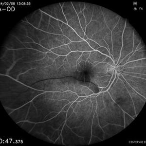
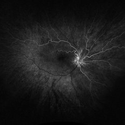
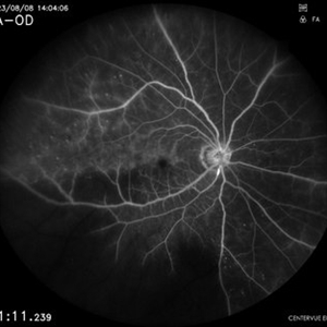 Branch Retinal Artery Occlusion
Mar 6 2024 by Akansha Sharma
Fluorescein angiography of a 67 year old male with capillary non perfusion areas in early phase in a case of branch retinal artery occlusion.
Photographer: Dr. Akansha Sharma, Bharati Eye Hospital
Condition/keywords: branch retinal artery occlusion, BRAO
-
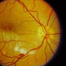
 Branch Retinal Artery Occlusion
Mar 6 2024 by Akansha Sharma
Color fundus photograph of a 67 year old male with branch retinal artery occlusion.
Photographer: Dr. Akansha Sharma, Bharati Eye Hospital
Condition/keywords: branch retinal artery occlusion, BRAO
-
 Branch Retinal Artery Occlusion
Mar 6 2024 by Akansha Sharma
Fluorescein angiography of a 65 year old male with absence of filling in the retinal artery close to the arcade in the early phase.
Photographer: Dr. Akansha Sharma, Bharati Eye Hospital
Condition/keywords: BRAO
-
 Branch Retinal Artery Occlusion
Mar 6 2024 by Akansha Sharma
Color fundus photograph of a 65 year old male with sudden dimness of vision presenting with a branch retinal artery occlusion.
Photographer: Dr. Akansha Sharma, Bharati Eye Hospital
Condition/keywords: branch retinal artery occlusion, BRAO
-
 Neovascularization at the Disc
Nov 10 2023 by Philip Conkling, MD
Fluorescein angiogram of a patient with a history of a branch retinal artery occlusion who developed neovascularization at the disc.
Condition/keywords: branch retinal artery occlusion (BRAO), Neovascularisation at the Disc (NVD)
-
 Neovascularization at the Disc
Nov 10 2023 by Philip Conkling, MD
Fluorescein angiogram of a patient with a history of a branch retinal artery occlusion who developed neovascularization at the disc.
Condition/keywords: branch retinal artery occlusion (BRAO), Neovascularisation at the Disc (NVD)
-
 Branch Retinal Artery Occlusion (BRAO)
Sep 26 2023 by Ben Serar
Fundus photograph of LE showing retinal edema and opacification along the superotemporal arcade, with cherry red spot at the macula, in a case of Branch Retinal Artery Occlusion (BRAO).
Condition/keywords: branch retinal artery occlusion (BRAO), cherry red spot
-
 Branch Retinal Artery Occlusion (BRAO)
Sep 21 2023 by Ben Serar
Fundus photograph of RE showing retinal edema and opacification along the inferotemporal vessel arcade, with cotton wool spots and flame shaped haemorrhage, in a case of Branch Retinal Artery Occlusion (BRAO).
Condition/keywords: branch retinal artery occlusion (BRAO)
-
 Branch Retinal Artery Occlusion (BRAO)
Sep 12 2023 by Ben Serar
Fundus photograph of the LE showing arterial occlusion along the inferotemporal vessel arcade with surrounding retinal edema and cotton-wool spots, in a case of Branch Retinal Artery Occlusion (BRAO).
Condition/keywords: branch retinal artery occlusion (BRAO), cotton wool spots, retinal edema
-
 BRAO with Hollenhorst plaque
Jun 27 2023 by Carlos Iván Campos Wolter, MD
Color fundus image of apatient with inferior branch retinal artery occlusion and a prominent Hollenhorst plaque
Photographer: Erika, Hospital Fundación Nuestra Señora de la Luz
Imaging device: TRC-NW8
Condition/keywords: branch retinal artery occlusion
-
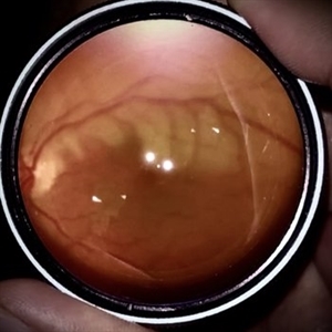
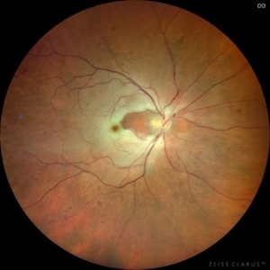 BRAO (Branch Retinal Artery Occlusion)
Jun 24 2023 by Mauricio Galvan Chavez, MD
Fundus photograph of a BRAO (Branch Retinal Artery Occlusion)
Photographer: Mauricio Galvan, Clinica de Retina, Guadalajara Jalisco.
Imaging device: Zeiss Clarus 700
Condition/keywords: arterial occlusion, branch retinal artery occlusion (BRAO), BRAO
-
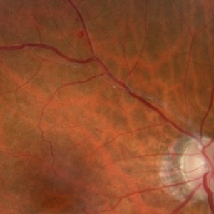
 Branch retinal artery occlusion
Jan 24 2023 by Rayna Marshall
Widefield fundus autofluorescence image of a 54-year-old female with an asymptomatic chronic branch retinal artery occlusion in the left eye. Hyper-autofluorescent embolus present at proximal inferior arcade, hypo-autoflorescence temporally corresponding to hyper-pigmentation. Vision was 20/20.
Photographer: Drew H. Scoles, MD, PhD, University of Pennsylvania
Condition/keywords: branch retinal artery occlusion (BRAO), BRAO, embolus
-
 Branch retinal artery occlusion
Jan 24 2023 by Rayna Marshall
Widefield fundus image of a 54-year-old female with an asymptomatic chronic branch retinal artery occlusion in the left eye. Peripheral schisis-like changes with pigmentation and temporal dot-blot hemorrhages. Vision was 20/20.
Photographer: Drew H. Scoles, MD, PhD, University of Pennsylvania
Condition/keywords: branch retinal artery occlusion (BRAO), BRAO, embolus
 Loading…