Search results (185 results)
-
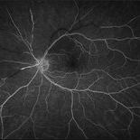
 Acute and Chronic OCT of BRAO
Sep 9 2021 by Aleksandra V. Rachitskaya, MD, FASRS
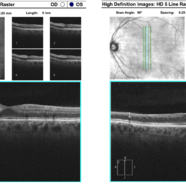
Acute and chronic OCT findings in BRAO. Acutely, inner retinal hyper-reflectivity is seen. Chronically, retina atrophy ensues.
Condition/keywords: BRAO, OCT
-
 Branch Retinal Artery Occlusion
Dec 15 2022 by Christopher R. Adam, M.D.
AF
Condition/keywords: branch retinal artery occlusion (BRAO), BRAO
-
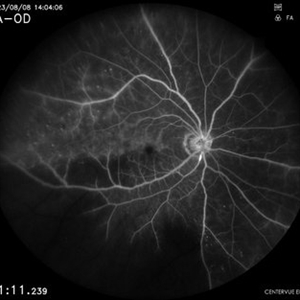
 Branch Retinal Artery Occlusion
Dec 15 2022 by Christopher R. Adam, M.D.
FA 32 seconds
Condition/keywords: branch retinal artery occlusion (BRAO), BRAO
-
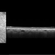
 Branch Retinal Artery Occlusion
Dec 15 2022 by Christopher R. Adam, M.D.
OCT
Condition/keywords: branch retinal artery occlusion (BRAO), BRAO
-
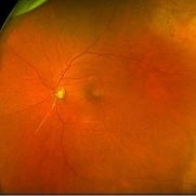
 Branch Retinal Artery Occlusion
Mar 6 2024 by Akansha Sharma
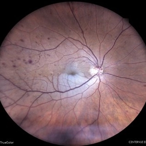
Color fundus photograph of a 65 year old male with sudden dimness of vision presenting with a branch retinal artery occlusion.
Photographer: Dr. Akansha Sharma, Bharati Eye Hospital
Condition/keywords: branch retinal artery occlusion, BRAO
-
 Branch Retinal Artery Occlusion
Mar 6 2024 by Akansha Sharma
Fluorescein angiography of a 65 year old male with absence of filling in the retinal artery close to the arcade in the early phase.
Photographer: Dr. Akansha Sharma, Bharati Eye Hospital
Condition/keywords: BRAO
-
 Branch Retinal Artery Occlusion
Mar 6 2024 by Akansha Sharma
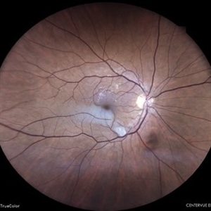
Color fundus photograph of a 67 year old male with branch retinal artery occlusion.
Photographer: Dr. Akansha Sharma, Bharati Eye Hospital
Condition/keywords: branch retinal artery occlusion, BRAO
-
 Branch Retinal Artery Occlusion
Mar 6 2024 by Akansha Sharma
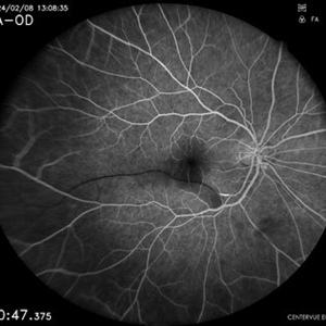
Fluorescein angiography of a 67 year old male with capillary non perfusion areas in early phase in a case of branch retinal artery occlusion.
Photographer: Dr. Akansha Sharma, Bharati Eye Hospital
Condition/keywords: branch retinal artery occlusion, BRAO
-
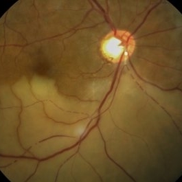
 Branch retinal artery occlusion
Jan 24 2023 by Rayna Marshall
OCT image of a 54-year-old female with an asymptomatic chronic branch retinal artery occlusion in the left eye showing inner retinal atrophy in the inferior macula corresponding to the region of chronic ischemia. Vision was 20/20.
Photographer: Drew H. Scoles, MD, PhD, University of Pennsylvania
Condition/keywords: branch retinal artery occlusion (BRAO), BRAO, embolus
-
 Branch retinal artery occlusion
Jan 24 2023 by Rayna Marshall
Widefield fundus image of a 54-year-old female with an asymptomatic chronic branch retinal artery occlusion in the left eye. Peripheral schisis-like changes with pigmentation and temporal dot-blot hemorrhages. Vision was 20/20.
Photographer: Drew H. Scoles, MD, PhD, University of Pennsylvania
Condition/keywords: branch retinal artery occlusion (BRAO), BRAO, embolus
-
 Branch retinal artery occlusion
Jan 24 2023 by Rayna Marshall
Widefield fundus autofluorescence image of a 54-year-old female with an asymptomatic chronic branch retinal artery occlusion in the left eye. Hyper-autofluorescent embolus present at proximal inferior arcade, hypo-autoflorescence temporally corresponding to hyper-pigmentation. Vision was 20/20.
Photographer: Drew H. Scoles, MD, PhD, University of Pennsylvania
Condition/keywords: branch retinal artery occlusion (BRAO), BRAO, embolus
-
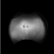
 BRAO
BRAO
Sep 10 2019 by Jason Griffith
59-year-old male referred with new onset loss of peripheral vision OS with cloudiness.
Photographer: Jodi Schiele, Tennessee Retina, Nashville, TN
Imaging device: Optos
Condition/keywords: branch retinal artery occlusion (BRAO)
-
 BRAO
BRAO
Oct 8 2012 by David R. Chow, MD, FRCS(C)
-
 BRAO
BRAO
Jun 29 2014 by John S. King, MD
BRAO.
Photographer: Wayne A Ladlee Jr
Condition/keywords: branch retinal artery occlusion (BRAO), embolic
-
 BRAO
BRAO
Jun 29 2014 by John S. King, MD
Deceased arterial flow in laminar phase of FA.
Photographer: Wayne A Ladlee Jr
Condition/keywords: branch retinal artery occlusion (BRAO), embolic
-
 BRAO
BRAO
Jun 29 2014 by John S. King, MD
AF with signal that may indicate calcific component of embolus.
Photographer: Wayne A Ladlee Jr
Condition/keywords: branch retinal artery occlusion (BRAO), embolic
-
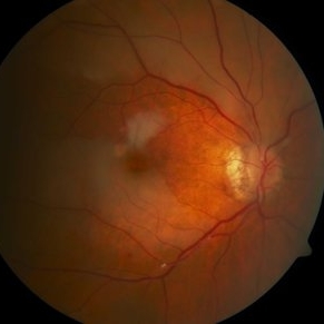
 BRAO (Branch Retinal Artery Occlusion)
Jun 24 2023 by Mauricio Galvan Chavez, MD
Fundus photograph of a BRAO (Branch Retinal Artery Occlusion)
Photographer: Mauricio Galvan, Clinica de Retina, Guadalajara Jalisco.
Imaging device: Zeiss Clarus 700
Condition/keywords: arterial occlusion, branch retinal artery occlusion (BRAO), BRAO
-
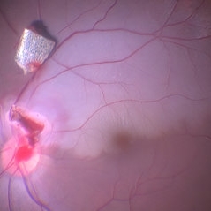 Intraocular Foreign Body with BRAO
Jan 12 2022 by Manish Nagpal, MD, FRCS (UK), FASRS
Intraoperative photo of a foreign body which was impacted on the edge of the disc leading to a BRAO. On table, the IOFB was loosened from the impact site and this photo was taken just prior to removal of the same using a magnet.
Photographer: Manish Nagpal, Director, Retina Foundation, Ahmedabad
Imaging device: Sony PMW -10 MD surgical camera
Condition/keywords: branch retinal artery occlusion (BRAO), BRAO, intraocular foreign body
-
 Superior Hemi-Central Retinal Artery Occlusion
Apr 24 2024 by Mosab Salah
Fundus photograph -inverted view- taken by smartphone fundus photography, of a young man with sudden onset altitudinal field defect, a Superior Hemi-Central Retinal Artery Occlusion noted.
Photographer: Dr Mosab Salah, The Islamic Hospital, Amman, Jordan
Imaging device: smartphone fundus photography and 30 D Lens
Condition/keywords: arterial occlusion, branch retinal artery occlusion (BRAO), BRAO, CRAO, Hemi-Central Retinal Artery Occlusion (CRAO), occlusive vasculitis, smartphone fundus photography
-
 BRAO with Hollenhorst plaque
Jul 22 2021 by Vishal Gupta, MBBS, MS
Fundus image of 54-year-old male patient with inferior branch retinal artery occlusion and a prominent Hollenhorst plaque seen as shining white dot at disk along with cattle trucking phenomenon.
Photographer: Dr Vishal Gupta, INHS Asvini, Mumbai, INDIA
Imaging device: Zeiss
Condition/keywords: branch retinal artery occlusion (BRAO), hollenhorst plaque
-
 BRAO d/t cat scratch disease - FA 00:18 min.
Jan 2 2013 by Roy Schwartz, MD
A 38-year-old male complained of a grey spot in visual field in his left eye. On clinical exam BRAO in LE, confirmed by FA, as seen in picture. Image shows delayed filling of artery. Serology for bartonella was positive.
Photographer: Galit Yair-Pur
Condition/keywords: branch retinal artery occlusion (BRAO), cat scratch retinitis
-
 BRAO d/t cat scratch disease - FA 5:23 min.
Jan 2 2013 by Roy Schwartz, MD
A 38-year-old male complained of a grey spot in visual field in his left eye. On clinical exam BRAO in LE, confirmed by FA, as seen in picture. The image was taken after late filling of artery. CWS blocks proximal part of artery. Serology for bartonella was positive.
Photographer: Galit Yair-Pur
Condition/keywords: branch retinal artery occlusion (BRAO), cat scratch retinitis
-
 BRAO d/t cat scratch disease - LE fundus photograph
Jan 2 2013 by Roy Schwartz, MD
A 38 year-old-male complained of a grey spot in visual field in his left eye. On clinical exam BRAO in LE, as seen in picture. Serology for bartonella was positive.
Photographer: Galit Yair-Pur
Condition/keywords: branch retinal artery occlusion (BRAO), cat scratch retinitis
-
 BRAO d/t cat scratch disease - RE fundus photograph
Jan 2 2013 by Roy Schwartz, MD
A 38-year-old male complained of a grey spot in visual field in his left eye. On clinical exam BRAO in LE, and a CWS in RE, as seen in this photograph. Serology for bartonella was positive.
Photographer: Galit Yair-Pur
Condition/keywords: branch retinal artery occlusion (BRAO), cat scratch retinitis
-
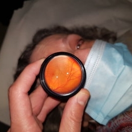 BRAO Indirect
Jan 4 2021 by Evgeny Gelman, MD
70-year-old healthy woman with 3 hours of "cloud" in front of her RE.
Photographer: Evgeny Gelman, shaare zedek medical center Israel
Imaging device: Personal Phone with 25D lens
Condition/keywords: branch retinal artery occlusion (BRAO)
 Loading…