Search results (143 results)
-
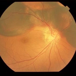
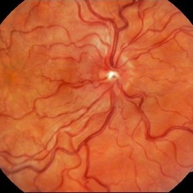 Retinal Aretriovenous Malformation
May 15 2014 by Mitzy E Torres Soriano, MD
Fundus photograph of a rare case of 18-year-old male with history of drug abuse. Visual acuity: hand motion. Specific diagnosis is unknown.
Photographer: Mitzy Torres Soriano. Hospital Central de Maracay. Venezuela
Imaging device: Zeiss, Inc
Condition/keywords: drug abuse, retinal arteriovenous malformations, vasculopathy
-
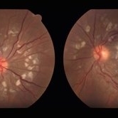 SLE Retinopathy
Nov 14 2016 by Mitzy E Torres Soriano, MD
25-year-old female patient with systemic lupus erythematosus. Photographs show cotton wool spots, intraretinal hemorrhages and vascular tortuosity. FA demonstrated retinal vasculitis and OCT revealed cystoid macular edema. In this case diagnosis of SLE was made after ocular manifestation.
Photographer: Grupo Laser Vision, Rosario, Argentina
Condition/keywords: cotton wool spots, occlusive retinal vasculitis, occlusive vasculitis, systemic lupus erythematosus, vasculopathy
-
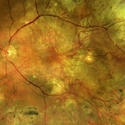
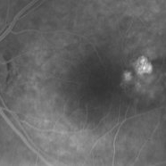
 Branching vascular network..Polypoidal choroidal vasculopathy
Feb 21 2022 by Shobhit Chawla, M.S.
A 56 year old patient on treatment with ANTIVGEF therapy for six years
Photographer: Shobhit Chawla
Imaging device: Zeiss Clarus 500
Condition/keywords: branching vascular network (BVN), polypoidal choroidal vasculopathy (PCV)
-
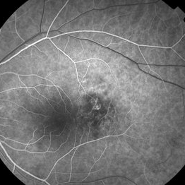
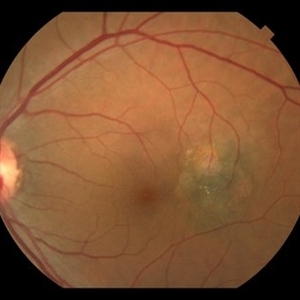
 BVN Branching Vascular Network in PCV
Feb 19 2022 by Vishal Gupta, MBBS, MS
Very evident branching vascular network in the fellow eye of one eyed patient who lost the other eye to massive hemorrhagic PCV in a 47 year old male patient.
Photographer: Dr Shobhit Chawla, Prakash Netra Kendr, Lucknow, UP, INDIA
Imaging device: Zeiss Clarus 500
Condition/keywords: branching vascular network (BVN), polypoidal choroidal vasculopathy (PCV)
-
 Choroidal Nevus with PCV
Jan 31 2018 by John S. King, MD
16 sec
Imaging device: topcon
Condition/keywords: choroidal neovascular membrane (CNVM), choroidal nevus, polypoidal choroidal vasculopathy (PCV)
-
 Choroidal Nevus with PCV
Jan 31 2018 by John S. King, MD
28 sec
Imaging device: topcon
Condition/keywords: choroidal neovascular membrane (CNVM), polypoidal choroidal vasculopathy (PCV)
-
 Choroidal Nevus with PCV
Jan 31 2018 by John S. King, MD
2:12
Imaging device: topcon
Condition/keywords: choroidal neovascular membrane (CNVM), choroidal nevus, polypoidal choroidal vasculopathy (PCV)
-
 Choroidal Nevus with PCV
Jan 31 2018 by John S. King, MD
44-year-old AAF without syptoms; nevus, rpe alterations, few exudates at base of polyp.
Imaging device: Topcon
Condition/keywords: choroidal neovascular membrane (CNVM), choroidal nevus, polypoidal choroidal vasculopathy (PCV)
-
 Choroidal nevus with polypoidal choroidal vasculopathy
Nov 20 2012 by Roy Schwartz, MD
Rare combination of a choroidal nevus complicated by polypoidal choroidal vasculopathy. The lesion is temporal to the fovea.
Photographer: Galit Yair-Pur
Condition/keywords: choroidal nevus, polypoidal choroidal vasculopathy (PCV)
-
 choroidal nevus with polypoidal choroidal vasculopathy
Nov 20 2012 by Roy Schwartz, MD
Rare combination of a choroidal nevus complicated by polypoidal choroidal vasculopathy. The lesion is temporal to the fovea, and leakage of subretinal fluid almost reaching the fovea is demonstrated.
Photographer: Galit Yair-Pur
Condition/keywords: choroidal nevus, polypoidal choroidal vasculopathy (PCV)
-
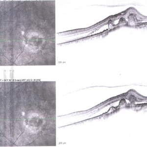 choroidal nevus with polypoidal choroidal vasculopathy
Nov 20 2012 by Roy Schwartz, MD
Rare combination of a choroidal nevus complicated by polypoidal choroidal vasculopathy. The lesion is temporal to the fovea, and subretinal fluid almost reaching the fovea is demonstrated.
Photographer: Galit Yair-Pur
Condition/keywords: choroidal nevus, optical coherence tomography (OCT), polypoidal choroidal vasculopathy (PCV)
-
 Disciform Scar
Aug 18 2020 by Aditya S Kelkar, MS, FRCS, FASRS,FRCOphth
Left eye fundus photograph of 75-year-old male, showing large disciform scar post subretinal bleeding secondary to idiopathic polypoidal choroidal vasculopathy
Photographer: Dr.Mounika Bolisetty
Imaging device: CLARUS 500
Condition/keywords: disciform scar, idiopathic polypoidal choroidal vasculopathy
-
 DISCIFORM SCAR AND RETINAL PIGMENT EPITHELIUM (RPE) DETACHMENT IN A CASE OF IDIOPATHIC POLYPOIDAL CHOROIDAL VASCULOPATHY (IPCV)
Oct 21 2023 by Aditya S Kelkar, MS, FRCS, FASRS,FRCOphth
Right eye fundus photograph of a 83 year old female demonstrating Disciform Scar And Retinal Pigment Epithelium (RPE) Detachment In A Case Of Idiopathic Polypoidal Choroidal Vasculopathy (IPCV).
Photographer: DR APURVA MUKADAM
Imaging device: OPTOS DAYTONA
Condition/keywords: disciform scar
-
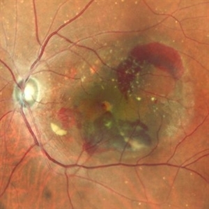
 Extra-Macula Subretinal Hemorrhage
Mar 21 2013 by Yusuke Oshima, MD, PhD
Fundus photograph of an 86-year-old woman with an extra-macula subretinal hemorrhage associated with polypoidal choroidal vasculopathy.
Photographer: Yusuke Takada, Osaka University Graduate School of Medicine
-
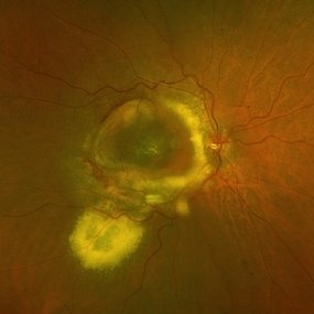
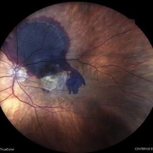
 Fundus Photo of IPCV
Aug 10 2019 by Manish Nagpal, MD, FRCS (UK), FASRS
Fundus showing serosanguinous collection under macular suggestive of active IPCV lesion.
Photographer: Gayathri Mohan, Retina Foundation
Imaging device: Nidek Mirante SLO
Condition/keywords: polypoidal choroidal vasculopathy (PCV)
-
---thumb.jpg/image-square;max$300,300.ImageHandler) Idiopathic Polypoidal Choroidal Vasculopathy
Idiopathic Polypoidal Choroidal Vasculopathy
Oct 18 2013 by Maurice F. Rabb
58 year old white male who initially presented to an ophthalmologist in November 1997 with decreased vision in his left eye. A diagnosis of idiopathic polypoidal choroidal vasculopathy was made.
Condition/keywords: idiopathic polypoidal choroidal vasculopathy
-
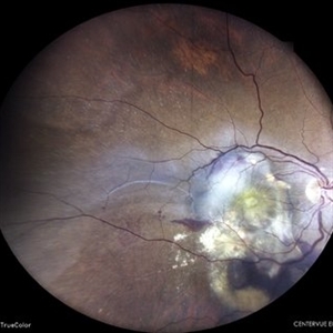
 Idiopathic Polypoidal Choroidal Vasculopathy
Jun 10 2020 by Manish Nagpal, MD, FRCS (UK), FASRS
Serosanguinous collection over macular area typical of IPCV.
Photographer: gaythri mohan
Imaging device: nidek slo mirante
Condition/keywords: idiopathic polypoidal choroidal vasculopathy
-
 IDIOPATHIC POLYPOIDAL CHOROIDAL VASCULOPATHY
Jun 6 2023 by Akansha Sharma
COLOUR FUNDUS PHOTOGRAPH OF AN 80 YEAR OLD MALE PATIENT WITH IDIOPATHIC POLYPOIDAL CHOROIDAL VASCULOPATHY
Photographer: Dr. Urmil Shah, Dr. Denish Patel, Dr. Akansha Sharma
Condition/keywords: idiopathic polypoidal choroidal vasculopathy, polypoidal choroidal vasculopathy (PCV)
-
 Idiopathic Polypoidal Choroidal Vasculopathy
Mar 26 2024 by Akansha Sharma
Color fundus photograph of a 61 year old treatment naive female patient with scarring at fovea with surrounding subretinal bleed suggestive of idiopathic polypoidal choroidal vasculopathy.
Photographer: Dr. Akansha Sharma, Bharati Eye Hospital
Condition/keywords: polypoidal choroidal vasculopathy (PCV)
 Loading…