Search results (143 results)
-
 Polypoidal Choroidal Vasculopathy-OCT
Aug 27 2012 by Young Hee Yoon, MD, PhD
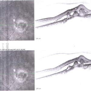
SD-OCT image of a 56-year-old woman. Her best-corrected visual acuity was 20/30.
Photographer: Kyoung Ree Kim, Asan Medical Center
Imaging device: Heidelberg Spectralis
Condition/keywords: polypoidal choroidal vasculopathy (PCV)
-
 Polypoidal Choroidal Vasculopathy
Aug 25 2012 by Hamid Ahmadieh, MD
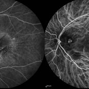
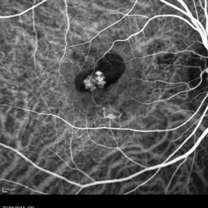
FA & ICG angiography imagings of a 73-year-old man with a peripapillary PCV.
Photographer: Hamid Ahmadieh, Ophthalmic Research Center, Labbafinejad Medical Center
Imaging device: Heidelberg Spectralis
Condition/keywords: indocyanine green (ICG) angiography, polypoidal choroidal vasculopathy (PCV)
-
 Polypoidal Choroidal Vasculopathy-FA
Aug 27 2012 by Young Hee Yoon, MD, PhD
Fluorescein Angiography(FA) image of a 56-year-old woman. Her best-corrected visual acuity was 20/30.
Photographer: Soo Hyun Cho, Asan Medical Center
Imaging device: Heidelberg
Condition/keywords: polypoidal choroidal vasculopathy (PCV)
-
 Polypoidal Choroidal Vasculopathy (ICG + OCT EDI)
May 16 2014 by Avris Romario Diparaja Siahaan
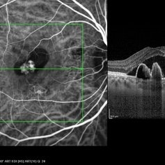
ICG + OCT (with EDI) (simultaneously)of a 46-year-old man with polypoidal choroidal vasculopathy (PCV).
Photographer: Avris Romario Diparaja Siahaan, Klinik Mata Nusantara
Imaging device: Heidelberg HRA + OCT Spectralis
Condition/keywords: optical coherence tomography (OCT), polypoidal choroidal vasculopathy (PCV)
-
 Polypoidal Choroidal Vasculopathy- Fundus photo
Aug 27 2012 by Young Hee Yoon, MD, PhD
Fundus photograph of a 56-year-old woman. Her best-corrected visual acuity was 20/30.
Photographer: Soon Tae Kim, Asan Medical Center
Imaging device: Canon
Condition/keywords: polypoidal choroidal vasculopathy (PCV)
-
 SLE Retinopathy
Nov 14 2016 by Mitzy E Torres Soriano, MD
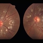
25-year-old female patient with systemic lupus erythematosus. Photographs show cotton wool spots, intraretinal hemorrhages and vascular tortuosity. FA demonstrated retinal vasculitis and OCT revealed cystoid macular edema. In this case diagnosis of SLE was made after ocular manifestation.
Photographer: Grupo Laser Vision, Rosario, Argentina
Condition/keywords: cotton wool spots, occlusive retinal vasculitis, occlusive vasculitis, systemic lupus erythematosus, vasculopathy
-
 Retinal Vasculopathy With Retinal Vasculitis and Ischemia
Jul 23 2014 by John S. King, MD
Purtscher's-like retinopathy.
Photographer: UPMC
Condition/keywords: lupus, systemic lupus erythematosus (SLE) retinopathy, systemic lupus erythematosus (SLE) vasculitis
-
 Polypoidal Choroidal Vasculopathy-ICG
Aug 27 2012 by Young Hee Yoon, MD, PhD
Indocyanine Green Angiography (ICGA) image of a 56-year-old woman. Her best-corrected visual acuity was 20/30.
Photographer: Kyoung Woon Kim, Asan Medical Center
Imaging device: Heidelberg
-
---thumb.jpg/image-square;max$300,300.ImageHandler) Idiopathic Polypoidal Choroidal Vasculopathy
Idiopathic Polypoidal Choroidal Vasculopathy
Oct 18 2013 by Maurice F. Rabb
58 year old white male who initially presented to an ophthalmologist in November 1997 with decreased vision in his left eye. A diagnosis of idiopathic polypoidal choroidal vasculopathy was made.
Condition/keywords: idiopathic polypoidal choroidal vasculopathy
-
 Polypoidal Choroidal Vasculopathy: Case 1 - Image 6of 7
Oct 4 2012 by Gregg T. Kokame, MD, MMM, FASRS
OCT/Indocyanine Green Angiography image of a 57-year-old woman with treatment-naive polypoidal choroidal vasculopathy. Series of images provides an comparative view of the same condition while utilizing a variet of different imaging procedures.
Photographer: Andrew Yuen, Retina Consultants of Hawaii
Imaging device: Heidelberg Spectralis
Condition/keywords: branching vascular network (BVN), indocyanine green (ICG) angiography, optical coherence tomography (OCT), polypoidal choroidal vasculopathy (PCV)
-
 choroidal nevus with polypoidal choroidal vasculopathy
Nov 20 2012 by Roy Schwartz, MD
Rare combination of a choroidal nevus complicated by polypoidal choroidal vasculopathy. The lesion is temporal to the fovea, and subretinal fluid almost reaching the fovea is demonstrated.
Photographer: Galit Yair-Pur
Condition/keywords: choroidal nevus, optical coherence tomography (OCT), polypoidal choroidal vasculopathy (PCV)
-
 Polypoidal Choroidal Vasculopathy: Case 1 - Image 4 of 7
Oct 4 2012 by Gregg T. Kokame, MD, MMM, FASRS
OCT/Indocyanine Green Angiography image of a 57-year-old woman with treatment-naive polypoidal choroidal vasculopathy. Series of images provides an comparative view of the same condition while utilizing a variet of different imaging procedures.
Photographer: Andrew Yuen, Retina Consultants of Hawaii
Imaging device: Heidelberg Spectralis
Condition/keywords: indocyanine green (ICG) angiography, optical coherence tomography (OCT), polypoidal choroidal vasculopathy (PCV)
-
 Pneumatic Displacement of a Massive Submacular Hemorrhage
Aug 3 2013 by Yusuke Oshima, MD, PhD
Pneumatic displacement of massive submacular hemorrhage with C3F8 gas.
Condition/keywords: gas pneumatic displacement, polypoidal choroidal vasculopathy (PCV), submacular hemorrhage, subretinal hemorrhage
-
---thumb.jpg/image-square;max$300,300.ImageHandler) Polypoidal Choroidal Vasculopathy: Case 1 - Image 2 of 7
Polypoidal Choroidal Vasculopathy: Case 1 - Image 2 of 7
Oct 4 2012 by Gregg T. Kokame, MD, MMM, FASRS
Bluepeak Autofluorescence image of a 57-year-old woman with treatment-naive polypoidal choroidal vasculopathy. Series of images provides an comparative view of the same condition while utilizing a variet of different imaging procedures.
Photographer: Andrew Yuen, Retina Consultants of Hawaii
Imaging device: Heidelberg Spectralis
Condition/keywords: autofluorescence imaging, fundus autofluorescence (FAF), polypoidal choroidal vasculopathy (PCV)
-
---thumb.jpg/image-square;max$300,300.ImageHandler) Polypoidal Choroidal Vasculopathy - Case 1
Polypoidal Choroidal Vasculopathy - Case 1
Oct 4 2012 by Gregg T. Kokame, MD, MMM, FASRS
Indocyanine Green Angiography image of a 57-year-old woman with treatment-naive polypoidal choroidal vasculopathy. Series of images provides an comparative view of the same condition while utilizing a variet of different imaging procedures.
Photographer: Andrew Yuen, Retina Consultants of Hawaii
Imaging device: Heidelberg Spectralis
Condition/keywords: branching vascular network (BVN), indocyanine green (ICG) angiography, polypoidal choroidal vasculopathy (PCV)
-
---thumb.jpg/image-square;max$300,300.ImageHandler) Polypoidal Choroidal Vasculopathy: Case 1 - Image 1 of 7
Polypoidal Choroidal Vasculopathy: Case 1 - Image 1 of 7
Oct 4 2012 by Gregg T. Kokame, MD, MMM, FASRS
Fluorescein Angiography image of a 57-year-old woman with treatment-naive polypoidal choroidal vasculopathy. Series of images provides an comparative view of the same condition while utilizing a variet of different imaging procedures.
Photographer: Andrew Yuen, Retina Consultants of Hawaii
Imaging device: Heidelberg Spectralis
Condition/keywords: fluorescein leakage, polypoidal choroidal vasculopathy (PCV)
-
---thumb.jpg/image-square;max$300,300.ImageHandler) Polypoidal Choroidal Vasculopathy
Polypoidal Choroidal Vasculopathy
Jul 13 2013 by Hamid Ahmadieh, MD
Late phase FA and ICG images of the right eye of a 55-year-old woman with decreased vision and metamorphopsia due to PCV.
Photographer: Elham Salehi, Negah Eye Center, Tehran
Imaging device: Heidelberg Spectralis
Condition/keywords: indocyanine green (ICG) angiography, polypoidal choroidal vasculopathy (PCV)
-
 choroidal nevus with polypoidal choroidal vasculopathy
Nov 20 2012 by Roy Schwartz, MD
Rare combination of a choroidal nevus complicated by polypoidal choroidal vasculopathy. The lesion is temporal to the fovea, and leakage of subretinal fluid almost reaching the fovea is demonstrated.
Photographer: Galit Yair-Pur
Condition/keywords: choroidal nevus, polypoidal choroidal vasculopathy (PCV)
-
 Polypoidal Choroidal Vasculopathy: Case 1 - Image 5 of 7
Oct 4 2012 by Gregg T. Kokame, MD, MMM, FASRS
OCT/Indocyanine Green Angiography image of a 57-year-old woman with treatment-naive polypoidal choroidal vasculopathy. Series of images provides an comparative view of the same condition while utilizing a variet of different imaging procedures.
Photographer: Andrew Yuen, Retina Consultants of Hawaii
Imaging device: Heidelberg Spectralis
Condition/keywords: indocyanine green (ICG) angiography, optical coherence tomography (OCT), polypoidal choroidal vasculopathy (PCV)
-
 Polypoidal Choroidal Vasculopathy: Case 1 - Image 7 of 7
Oct 4 2012 by Gregg T. Kokame, MD, MMM, FASRS
OCT/Indocyanine Green Angiography image of a 57-year-old woman with treatment-naive polypoidal choroidal vasculopathy. Series of images provides an comparative view of the same condition while utilizing a variet of different imaging procedures.
Photographer: Andrew Yuen, Retina Consultants of Hawaii
Imaging device: Heidelberg Spectralis
Condition/keywords: branching vascular network (BVN), indocyanine green (ICG) angiography, optical coherence tomography (OCT), polypoidal choroidal vasculopathy (PCV)
-
---thumb.jpg/image-square;max$300,300.ImageHandler) Idiopathic Polypoidal Choroidal Vasculopathy
Idiopathic Polypoidal Choroidal Vasculopathy
Oct 18 2013 by Maurice F. Rabb
58 year old white male who initially presented to an ophthalmologist in November 1997 with decreased vision in his left eye. A diagnosis of idiopathic polypoidal choroidal vasculopathy was made.
Condition/keywords: idiopathic polypoidal choroidal vasculopathy
-
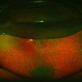
 Polypoidal Choroidal Vasculopathy
Feb 8 2013 by Sjakon G Tahija, MD
Fundus photograph of a 40-year-old woman who suffered from polypoidal choroidal vasculopathy (PCV). She had an injection of intravitreal Avastin and shortly afterwards had breakthrough hemorrhage.
Photographer: Arvis Siahaan, Klinik Mata Nusantara, Jakarta, Indonesia
Imaging device: Topcon TRC 50DX Type IA
Condition/keywords: polypoidal choroidal vasculopathy (PCV)
-
---thumb.jpg/image-square;max$300,300.ImageHandler) Polypoidal Choroidal Vasculopathy: Case 1 - Image 3 of 7
Polypoidal Choroidal Vasculopathy: Case 1 - Image 3 of 7
Oct 4 2012 by Gregg T. Kokame, MD, MMM, FASRS
Fluorescein Angriography image of a 57-year-old woman with treatment-naive polypoidal choroidal vasculopathy. Series of images provides an comparative view of the same condition while utilizing a variet of different imaging procedures.
Photographer: Andrew Yuen, Retina Consultants of Hawaii
Imaging device: Heidelberg Spectralis
Condition/keywords: infrared image, polypoidal choroidal vasculopathy (PCV)
-
 SLE Retinal Vasculopathy With Retinal Vasculitis and Ischemia
Jul 23 2014 by John S. King, MD
OD: 45 sec and 3 min OS: 1:30 min and 3 min
Photographer: UPMC
Condition/keywords: lupus, systemic lupus erythematosus (SLE) retinopathy, systemic lupus erythematosus (SLE) vasculitis
-
 Polypoidal Choroidal Vasculopathy (ICG Early Phase)
May 16 2014 by Avris Romario Diparaja Siahaan
ICG angiography image of a 46-year-old man with polypoidal choroidal vasculopathy (PCV).
Photographer: Avris Romario Diparaja Siahaan, Klinik Mata Nusantara
Imaging device: Heidelberg HRA + OCT Spectralis
Condition/keywords: indocyanine green (ICG) angiography, polypoidal choroidal vasculopathy (PCV)
 Loading…