Search results (143 results)
-
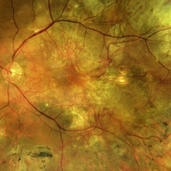
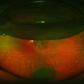
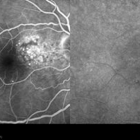
 Disciform Scar
Aug 18 2020 by Aditya S Kelkar, MS, FRCS, FASRS,FRCOphth
Left eye fundus photograph of 75-year-old male, showing large disciform scar post subretinal bleeding secondary to idiopathic polypoidal choroidal vasculopathy
Photographer: Dr.Mounika Bolisetty
Imaging device: CLARUS 500
Condition/keywords: disciform scar, idiopathic polypoidal choroidal vasculopathy
-
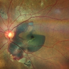
 BVN Branching Vascular Network in PCV
Feb 19 2022 by Vishal Gupta, MBBS, MS
Very evident branching vascular network in the fellow eye of one eyed patient who lost the other eye to massive hemorrhagic PCV in a 47 year old male patient.
Photographer: Dr Shobhit Chawla, Prakash Netra Kendr, Lucknow, UP, INDIA
Imaging device: Zeiss Clarus 500
Condition/keywords: branching vascular network (BVN), polypoidal choroidal vasculopathy (PCV)
-
 choroidal nevus with polypoidal choroidal vasculopathy
Nov 20 2012 by Roy Schwartz, MD
Rare combination of a choroidal nevus complicated by polypoidal choroidal vasculopathy. The lesion is temporal to the fovea, and leakage of subretinal fluid almost reaching the fovea is demonstrated.
Photographer: Galit Yair-Pur
Condition/keywords: choroidal nevus, polypoidal choroidal vasculopathy (PCV)
-
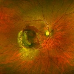
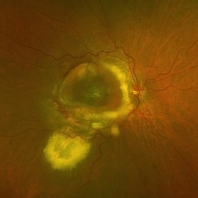 DISCIFORM SCAR AND RETINAL PIGMENT EPITHELIUM (RPE) DETACHMENT IN A CASE OF IDIOPATHIC POLYPOIDAL CHOROIDAL VASCULOPATHY (IPCV)
Oct 21 2023 by Aditya S Kelkar, MS, FRCS, FASRS,FRCOphth
Right eye fundus photograph of a 83 year old female demonstrating Disciform Scar And Retinal Pigment Epithelium (RPE) Detachment In A Case Of Idiopathic Polypoidal Choroidal Vasculopathy (IPCV).
Photographer: DR APURVA MUKADAM
Imaging device: OPTOS DAYTONA
Condition/keywords: disciform scar
-
 Fundus Photo of IPCV
Aug 10 2019 by Manish Nagpal, MD, FRCS (UK), FASRS
Fundus showing serosanguinous collection under macular suggestive of active IPCV lesion.
Photographer: Gayathri Mohan, Retina Foundation
Imaging device: Nidek Mirante SLO
Condition/keywords: polypoidal choroidal vasculopathy (PCV)
-
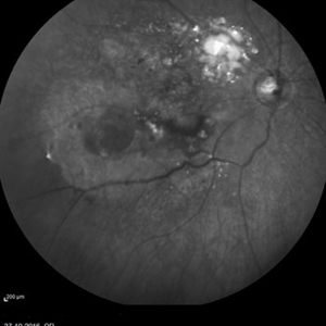 PCV
PCV
Nov 1 2016 by Apoorva Guruprasad Ayachit, MS
Infra red photograph of 72-year-old man with an area of exudation superotemporal to disc and powdery deposits. PED is seen inferotemporal to macula.
Photographer: Apoorva Ayachit
Imaging device: Heidelberg
Condition/keywords: polypoidal choroidal vasculopathy (PCV)
-
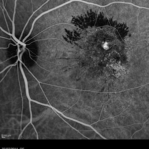 PCV
PCV
Jul 26 2014 by Avris Romario Diparaja Siahaan
An ICG image of a 65-year-old-woman with PCV on her left eye (mid phase).
Photographer: Avris Romario Diparaja Siahaan, Klinik Mata Nusantara
Imaging device: Heidelberg Spectralis
Condition/keywords: indocyanine green (ICG) angiography, polypoidal choroidal vasculopathy (PCV)
-
 Pneumatic Displacement of a Massive Submacular Hemorrhage
Aug 3 2013 by Yusuke Oshima, MD, PhD
Pneumatic displacement of massive submacular hemorrhage with C3F8 gas.
Condition/keywords: gas pneumatic displacement, polypoidal choroidal vasculopathy (PCV), submacular hemorrhage, subretinal hemorrhage
-
 Polyploidal Choroidal Vasculopathy
Dec 27 2024 by Tejaswita Verma
Fundus image of a 74 year old woman with CF1mt vision in right eye showing large PED in a case of PCV. There was associated full thickness macular hole in the same eye.
Photographer: DR. TEJASWITA VERMA
Imaging device: MIRANTE
Condition/keywords: PED, polypoidal choroidal vasculopathy (PCV)
-
 Polypoidal Choroidal Vasculopathy
Mar 13 2018 by Gabriel Costa Andrade, PhD
Fundus image of the right eye of a 76-year-old man with Polypoidal Choroidal Vasculopathy in the right eye.
Photographer: Gabriel Andrade, MD
Imaging device: Optos® California
Condition/keywords: neovascular age-related macular degeneration (AMD)
-
 Polypoidal Choroidal Vasculopathy-ICG
Aug 27 2012 by Young Hee Yoon, MD, PhD
Indocyanine Green Angiography (ICGA) image of a 56-year-old woman. Her best-corrected visual acuity was 20/30.
Photographer: Kyoung Woon Kim, Asan Medical Center
Imaging device: Heidelberg
-
 Submacular Hemorrhage PCV
May 6 2022 by Shobhit Chawla, M.S.
Submacular hemorrhage in a 38 years old female patient cause polyp bleed in PCV.
Photographer: Shobhit Chawla
Imaging device: Zeiss Clarus 500
Condition/keywords: polypoidal choroidal vasculopathy (PCV), submacular hemorrhage
-
 Submacular Hemorrhage Before Treatment
Mar 21 2013 by Yusuke Oshima, MD, PhD
Fundus photograph of an 83-year-old man with a submacular hemorrhage due to polypoidal choroidal vasculopathy.
Photographer: Yusuke Takada, Osaka University Graduate School of Medicine
Condition/keywords: submacular hemorrhage
-
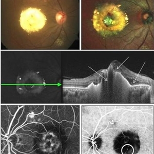
 Polypoidal Choroidal Vasculopathy: Case 1 - Image 6of 7
Oct 4 2012 by Gregg T. Kokame, MD, MMM, FASRS
OCT/Indocyanine Green Angiography image of a 57-year-old woman with treatment-naive polypoidal choroidal vasculopathy. Series of images provides an comparative view of the same condition while utilizing a variet of different imaging procedures.
Photographer: Andrew Yuen, Retina Consultants of Hawaii
Imaging device: Heidelberg Spectralis
Condition/keywords: branching vascular network (BVN), indocyanine green (ICG) angiography, optical coherence tomography (OCT), polypoidal choroidal vasculopathy (PCV)
-
 Polypoidal Choroidal Vasculopathy
Feb 8 2013 by Sjakon G Tahija, MD
Fundus photograph of a 40-year-old woman who suffered from polypoidal choroidal vasculopathy (PCV). She had an injection of intravitreal Avastin and shortly afterwards had breakthrough hemorrhage.
Photographer: Arvis Siahaan, Klinik Mata Nusantara, Jakarta, Indonesia
Imaging device: Topcon TRC 50DX Type IA
Condition/keywords: polypoidal choroidal vasculopathy (PCV)
-
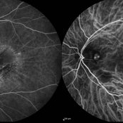
 Polypoidal Choroidal Vasculopathy - IVFA/ICGA
Jun 29 2018 by Gareth Lema, MD, PhD
IVFA and ICGA at 15 minutes.
Photographer: Sandra Boglione, Ross Eye Institute, University at Buffalo Jacobs School of Medicine, Buffalo, NY
Imaging device: Heidelberg
Condition/keywords: polypoidal choroidal vasculopathy (PCV)
-
---thumb.JPG/image-square;max$300,300.ImageHandler) Polypoidal Choroidal Vasculopathy with Massve Subretinal Hemorrhage
Polypoidal Choroidal Vasculopathy with Massve Subretinal Hemorrhage
Mar 9 2013 by Young-Gyun Kim, MD
Fundus photograph of a 52-year-old man with PCV and massive subretinal hemorrhage.
Photographer: Shin Ji-Young, Eulji university, Seoul
Imaging device: Topcon TRC 50 EX
Condition/keywords: polypoidal choroidal vasculopathy (PCV)
-
 Polypoidal Choroidal Vasculopathy-OCT
Aug 27 2012 by Young Hee Yoon, MD, PhD
SD-OCT image of a 56-year-old woman. Her best-corrected visual acuity was 20/30.
Photographer: Kyoung Ree Kim, Asan Medical Center
Imaging device: Heidelberg Spectralis
Condition/keywords: polypoidal choroidal vasculopathy (PCV)
-
 Polypoidal Chroidal Vasculopathy
Sep 21 2018 by Dhaivat Shah
A 40-year-old female presented with sudden onset decreased vision in right eye. BCVA: CF 1 mt. Fundus showed massive subretinal exudation with haemorrhage. EDI OCT showed notched PEDs with shallow SRF and exudation with back-shadowing. FFA shows leak with window defects. ICG shows hotspot in late phase. Polypoidal choroidal vasculopathy (PCV) is a retinal disorder characterized by the presence of aneurysmal polypoidal lesions in the choroidal vasculature, resulting in damage to the overlying retina and loss of retinal pigment epithelium. The aneurysmal dilatations, also known as polyps, may be found subfoveal, juxtafoveal, extrafoveal, peripapillary or even peripheral regions. The polypoidal lesions are best detected on indocyanine green angiography as hotspots in late phase. The presence of choroidal polyps can lead to recurrent episodes of exudative retinal detachment, serous or hemorrhagic pigment epithelial detachment, subretinal hemorrhage and exudation. Treatment is available in form of laser/PDT along with Anti VEGF injection.
Photographer: Miss Moupiya Das
Condition/keywords: polypoidal choroidal vasculopathy (PCV)
-
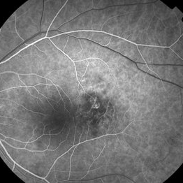
 Polypoidal Choroidal Vasculopathy
Aug 25 2012 by Hamid Ahmadieh, MD
FA & ICG angiography imagings of a 73-year-old man with a peripapillary PCV.
Photographer: Hamid Ahmadieh, Ophthalmic Research Center, Labbafinejad Medical Center
Imaging device: Heidelberg Spectralis
Condition/keywords: indocyanine green (ICG) angiography, polypoidal choroidal vasculopathy (PCV)
-
 Polypoidal Choroidal Vasculopathy- Fundus photo
Aug 27 2012 by Young Hee Yoon, MD, PhD
Fundus photograph of a 56-year-old woman. Her best-corrected visual acuity was 20/30.
Photographer: Soon Tae Kim, Asan Medical Center
Imaging device: Canon
Condition/keywords: polypoidal choroidal vasculopathy (PCV)
-
 Polypoidal Choroidal Vasculopathy
Aug 15 2015 by Thomas A. Ciulla, MD, MBA, FASRS
The patient underwent multiple antiVEGF treatments. Six months after initial presentation, the macular exam and OCT improved considerably.
Condition/keywords: idiopathic polypoidal choroidal vasculopathy
-
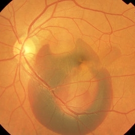
 Branching vascular network..Polypoidal choroidal vasculopathy
Feb 21 2022 by Shobhit Chawla, M.S.
A 56 year old patient on treatment with ANTIVGEF therapy for six years
Photographer: Shobhit Chawla
Imaging device: Zeiss Clarus 500
Condition/keywords: branching vascular network (BVN), polypoidal choroidal vasculopathy (PCV)
-
 Choroidal Nevus with PCV
Jan 31 2018 by John S. King, MD
16 sec
Imaging device: topcon
Condition/keywords: choroidal neovascular membrane (CNVM), choroidal nevus, polypoidal choroidal vasculopathy (PCV)
-
 Choroidal Nevus with PCV
Jan 31 2018 by John S. King, MD
28 sec
Imaging device: topcon
Condition/keywords: choroidal neovascular membrane (CNVM), polypoidal choroidal vasculopathy (PCV)
 Loading…