Search results (92 results)
-
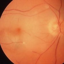
---thumb.jpg/image-square;max$300,300.ImageHandler) Acute retinal necrosis
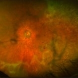
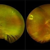
Acute retinal necrosis
Feb 15 2013 by From the Collections of Thomas M. Aaberg, MD and Thomas M. Aaberg Jr., MD
Diffuse intraretinal hemorrhages and whitening in the posterior pole consistent with acute retinal necrosis.
Condition/keywords: macular edema, microangiopathy, retinal necrosis, retinal whitening
-
 Central Retinal Artery Occlusion
Mar 26 2019 by Gary R. Cook, MD, FACS
61-year-old male patient with acute CRAO OS demonstrating a hyperemic optic disc with a couple of peripapillary hemorrhages, generalized arteriolar narrowing, a cherry-red spot in the macula, and retinal whitening surrounding the fovea; VA= LP.
Imaging device: Topcon VT-50
Condition/keywords: central retinal artery occlusion (CRAO), cherry red spot, retinal whitening
-
 Central Retinal Artery Occlusion with Cilioretinal Sparing
Mar 26 2019 by Gary R. Cook, MD, FACS
81-year-old Vietnamese female with acute CRAO with cilioretinal artery sparing OS; VA= 20/60.
Imaging device: Topcon VT-50
Condition/keywords: central retinal artery occlusion (CRAO), cilioretinal sparing, retinal whitening
-
 Cilioretinal Artery Occlusion
May 14 2024 by Eloy Mata-Cortes, MD
Color image capturing the left eye of a 32-year-old female. Despite a negative ophthalmological and medical history, she reported three days of blurred vision and a paracentral scotoma in her left eye, while maintaining central vision. The image reveals retinal whitening, extends from the parafoveal region to the inferotemporal arcade indicative of cilioretinal artery occlusion. Following this observation, the patient was referred for systemic assessment to explore the underlying etiology of the occlusion.
Photographer: Eloy Mata-Cortes, MD, Instituto Mexicano de Oftalmología, Querétaro, México
Imaging device: Nidek Mirante
Condition/keywords: cilioretinal artery occlusion, oclussion, retinal whitening
-
 Commotio-Retinae
Sep 22 2021 by Luiz Guilherme Freitas, MD, MsC, PhD
Fundus photograph of a 30-year-old male patient with blunt injury to the globe. Commotio retinae is retinal whitening/opacification that results from a blunt injury. The ocular findings will often resolve in a matter of days to weeks. Vision loss can result from commotio involving the posterior pole (historically referred to as Berlin’s edema). Clinical findings of commotio include the characteristic retinal whitening. Commotio may result in significant vision loss that can be transient. Healing can result in pigmentary changes and retinal thinning which may be associated with poor visual recovery if the area of involvement is macular.
Photographer: Diogo Melo, Santa Luzia Eye Hospital Recife - PE – Brazil
Condition/keywords: Berlin's edema, blunt trauma, commotio retinae, retinal whitening
-
 Embolic Central Retinal Artery Occlusion
Mar 26 2019 by Gary R. Cook, MD, FACS
58-year-old WM with embolic CRAO demonstrating a a cherry-red spot in macula, retinal whitening around the fovea, and the embolus in a inferotemporal branch retinal arteriole; VA= HM 6''
Imaging device: Topcon VT-50
Condition/keywords: central retinal artery occlusion (CRAO), cherry red spot, embolus, retinal whitening
-
 Progressive Outer Retinal Necrosis
Nov 30 2018 by Nichole Lewis
Fluorescein angiogram of an 86-year-old male with progressive outer retinal necrosis and chronic cystoid macular edema. This patient has occlusive vasculitis with non-perfusion, significant retinitis, retinal whitening and intra-retinal hemorrhages. Patient is immunosupressed with a history of kidney transplantation. VA 20/60. Patient was treated with intravitreal foscarnet and admitted to the hospital for an infectious disease and transplant team consultation.
Photographer: Nichole Lewis
Condition/keywords: cystoid macular edema (CME), intraretinal hemorrhage, non-perfusion, occlusive vasculitis, progressive outer retinal necrosis (PORN), retinal whitening, retinitis
-
 Progressive Outer Retinal Necrosis
Nov 30 2018 by Nichole Lewis
Fluorescein angiogram of an 86-year-old male with progressive outer retinal necrosis and chronic cystoid macular edema. This patient has occlusive vasculitis with non-perfusion, significant retinitis, retinal whitening and intra-retinal hemorrhages. Patient is immunosupressed with a history of kidney transplantation. VA 20/60. Patient was treated with intravitreal foscarnet and admitted to the hospital for an infectious disease and transplant team consultation.
Photographer: Nichole Lewis
Condition/keywords: cystoid macular edema (CME), intragel hemorrhage, non-perfusion, occlusive vasculitis, progressive outer retinal necrosis (PORN), retinal whitening, retinitis
-
 Progressive Outer Retinal Necrosis
Nov 5 2019 by Nichole Lewis
86-year-old male with progressive outer retinal necrosis, significant retinitis, retinal whitening, intraretinal hemorrhages and peripheral rpe changes. FA showed occlusive vasculitis with non-perfusion. Patient is immuno-suppressed with a history of renal transplant. VA 20/60.
Photographer: Nichole Lewis
Imaging device: Optos
Condition/keywords: intraretinal hemorrhage, occlusive vasculitis, progressive outer retinal necrosis (PORN), retinal pigment epithelium (RPE) changes, retinal whitening, retinitis
-
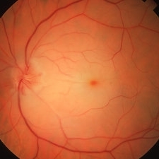
---thumb.jpg/image-square;max$300,300.ImageHandler) Retinal whitening consistent with ocular toxoplasmosis
Retinal whitening consistent with ocular toxoplasmosis
Feb 15 2013 by From the Collections of Thomas M. Aaberg, MD and Thomas M. Aaberg Jr., MD
Color fundus photograph showing retinal whitening consistent with ocular toxoplasmosis.
Condition/keywords: ocular toxoplasmosis
-
 Retinitis Sclopetaria
Jun 29 2018 by Gareth Lema, MD, PhD
Retinal whitening, subretinal hemorrhages, retinal hemorrhages, and vascular tortuosity following blunt trauma from a paintball.
Photographer: Flaum Eye Institute, University of Rochester, Rochester, NY
Condition/keywords: blunt trauma, chorioretinitis sclopetaria
-
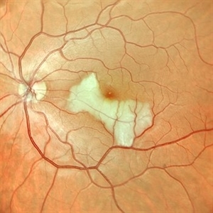
 Aborted Arteriolitis
Feb 15 2013 by From the Collections of Thomas M. Aaberg, MD and Thomas M. Aaberg Jr., MD
Fundus photograph showing activated toxoplasma retinochoroiditis with active retinal whitening adjacent to a hyperpigmented scar in the superonasal mid-periophery.
Condition/keywords: ocular toxoplasmosis
-
 Acute Central Retinal Artery Occlusion with Natural Reperfusion
Mar 12 2021 by Kushal S Delhiwala, MBBS, MS, FMRF,FICO, FAICO
Fundus photographs of 33-year-old healthy male with right eye acute CRAO of 12 hours duration showing cattle trucking, extensive retinal whitening and cherry red spot (left image). Right image 18 hours later showing reduced extent of retinal whitening and absent cattle trucking, suggestive of natural restoration of perfusion.
Photographer: Kushal Delhiwala, Netralaya superspeciality eye hospital, Ahmedabad, Gujarat,India
Imaging device: Optos Daytona
Condition/keywords: cattle trucking, central retinal artery occlusion (CRAO), cherry red spot
-
 Acute Compressive Optic Neuropathy
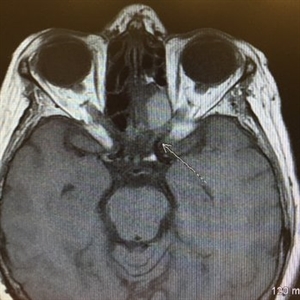
Jun 1 2019 by John S. King, MD
84-year-old white female with acute loss of vision in the left eye one day ago was sent here after going to the ED per primary eye provider. She described vision loss as a grey curtain that became total darkness. She had left sided temporal tenderness and some left sided neck pain. In the ED the cardiac work-up was u/r, the ESR and CRP were normal, and the CTH showed some non-specific opacification in the L ethmoid sinus. Acuity was HM OS with RAPD, normal EOMs, no proptosis or ptosis, posteriorly no SVPs were noted; the optic discs were pink and flat; no emboli or retinal whitening present; some bear tracks located nasally (see photo). She was referred to Dr. Doyle, who ordered an MRI, which showed a large mucocele with bony erosion into the left orbit, along with some ON enhancement possibly from compression (see images). She was operated that night and later recovered to 20/40 in that eye with a residual, inferior arcuate scotoma.
Condition/keywords: bear tracks, optic neuropathy
-
 Acute Optic Neuropathy Due to Large Mucocele
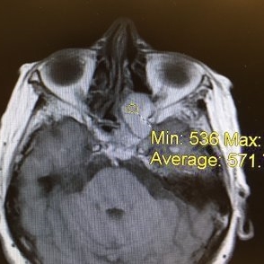
Jun 1 2019 by John S. King, MD
84-year-old white female with acute loss of vision in the left eye one day ago was sent here after going to the ED per primary eye provider. She described vision loss as a grey curtain that became total darkness. She had left sided temporal tenderness and some left sided neck pain. In the ED the cardiac work-up was u/r, the ESR and CRP were normal, and the CTH showed some non-specific opacification in the L ethmoid sinus. Acuity was HM OS with RAPD, normal EOMs, no proptosis or ptosis, posteriorly no SVPs were noted; the optic discs were pink and flat; no emboli or retinal whitening present; some bear tracks located nasally (see photo). She was referred to Dr. Doyle, who ordered an MRI, which showed a large mucocele with bony erosion into the left orbit, along with some ON enhancement possibly from compression (see Images). She was operated that night and later recovered to 20/40 in that eye with a residual, inferior arcuate scotoma.
Condition/keywords: bear tracks, optic neuropathy
-
 Acute Optic Neuropathy Due to Large Mucocele (Incidental Bear Tracks)
Jun 1 2019 by John S. King, MD
84-year-old white female with acute loss of vision in the left eye one day ago was sent here after going to the ED per primary eye provider. She described vision loss as a grey curtain that became total darkness. She had left sided temporal tenderness and some left sided neck pain. In the ED the cardiac work-up was u/r, the ESR and CRP were normal, and the CTH showed some non-specific opacification in the L ethmoid sinus. Acuity was HM OS with RAPD, normal EOMs, no proptosis or ptosis, posteriorly no SVPs were noted; the optic discs were pink and flat; no emboli or retinal whitening present; some bear tracks located nasally (see photo). She was referred to Dr. Doyle, who ordered an MRI, which showed a large mucocele with bony erosion into the left orbit, along with some ON enhancement possibly from compression (see images). She was operated that night and later recovered to 20/40 in that eye with a residual, inferior arcuate scotoma.
Photographer: Karin Aletter
Imaging device: Topcon 50
Condition/keywords: bear tracks, optic neuropathy
-
 ARN (#1) Initial Photo
May 27 2019 by John S. King, MD
60-year-old African American female who had been treated for iridocyclitis for at least a week sent in for vitritis and a nasal fundus lesion. Complaints included redness, floaters, photophobia, and decreased vision. Husband had recent shingles. Acuity was 20/60-2 with IOP of 12, and small KP in Art's triangel, 1-2+ a/c cell, 2-3+ ant vit cell, diffuse arteriolar sheathing, multiple areas of retinal whitening in periphery and mid-periphery (see Photo #1). PCR of a/c was performed, and intravitreal GCV administered, and VACV 2g qid and ASA started.... PCR positive for HZV, pred taper was started two days after presentation as the infection had begun to stablize..... Five days from presentation the vision was 20/60, inflammation and areas of retinal whitening had improved (see Photo #2).... One week later acuity was 20/30, the a/c was quiet and KP resolved; ant vitreous cell decreased; and there was further improvement in retinal appearance without any signs of retinal holes or detachment; she is now on low dose maint VACV (see photo#3)
Photographer: Maysee Yang
Imaging device: Optos CA
Condition/keywords: acute retinal necrosis, Herpes zoster
-
 ARN (#2) Five Days Since Initial Visit
May 27 2019 by John S. King, MD
60-year-old African American female who had been treated for iridocyclitis for at least a week sent in for vitritis and a nasal fundus lesion. Complaints included redness, floaters, photophobia, and decreased vision. Husband had recent shingles. Acuity was 20/60-2 with IOP of 12, and small KP in Art's triangel, 1-2+ a/c cell, 2-3+ ant vit cell, diffuse arteriolar sheathing, multiple areas of retinal whitening in periphery and mid-periphery (see Photo #1). PCR of a/c was performed, and intravitreal GCV administered, and VACV 2g qid and ASA started.... PCR positive for HZV, pred taper was started two days after presentation as the infection had begun to stablize..... Five days from presentation the vision was 20/60, inflammation and areas of retinal whitening had improved (see Photo #2).... One week later acuity was 20/30, the a/c was quiet and KP resolved; ant vitreous cell decreased; and there was further improvement in retinal appearance without any signs of retinal holes or detachment; she is now on low dose maint VACV (see photo#3)
Photographer: Maysee Yang
Imaging device: Optos CA
Condition/keywords: acute retinal necrosis, Herpes zoster
-
 ARN (#3) This is comparison between the latest visit (left) and one week prior (which is the right photo, and same one as photo #2)
May 27 2019 by John S. King, MD
60-year-old African American female who had been treated for iridocyclitis for at least a week sent in for vitritis and a nasal fundus lesion. Complaints included redness, floaters, photophobia, and decreased vision. Husband had recent shingles. Acuity was 20/60-2 with IOP of 12, and small KP in Art's triangel, 1-2+ a/c cell, 2-3+ ant vit cell, diffuse arteriolar sheathing, multiple areas of retinal whitening in periphery and mid-periphery (see Photo #1). PCR of a/c was performed, and intravitreal GCV administered, and VACV 2g qid and ASA started.... PCR positive for HZV, pred taper was started two days after presentation as the infection had begun to stablize..... Five days from presentation the vision was 20/60, inflammation and areas of retinal whitening had improved (see Photo #2).... One week later acuity was 20/30, the a/c was quiet and KP resolved; ant vitreous cell decreased; and there was further improvement in retinal appearance without any signs of retinal holes or detachment; she is now on low dose maint VACV (see photo#3)
Photographer: Maysee Yang
Imaging device: Optos CA
Condition/keywords: acute retinal necrosis, Herpes zoster
-
---thumb.jpg/image-square;max$300,300.ImageHandler) Behcet Uveitis
Behcet Uveitis
Feb 15 2013 by From the Collections of Thomas M. Aaberg, MD and Thomas M. Aaberg Jr., MD
Color fundus photographs of the right eye of a patient suspected to have Behcet Uveitis. Over the course of 11 days, there is progressive optic disc edema, intraretinal whitening, hemorrhage and vessel occlusion.
Condition/keywords: Behcet's uveitis, posterior uveitis, retinitis
-
---thumb.jpg/image-square;max$300,300.ImageHandler) Behcet Uveitis
Behcet Uveitis
Feb 15 2013 by From the Collections of Thomas M. Aaberg, MD and Thomas M. Aaberg Jr., MD
Color fundus photographs of the right eye of a patient suspected to have Behcet Uveitis. Over the course of 11 days, there is progressive optic disc edema, intraretinal whitening, hemorrhage and vessel occlusion. Fluorescein angiography confirms impaired retinal perfusion secondary to vessel occlusion.
Condition/keywords: posterior uveitis, retinitis
-
---thumb.jpg/image-square;max$300,300.ImageHandler) Behcet Uveitis.
Behcet Uveitis.
Feb 15 2013 by From the Collections of Thomas M. Aaberg, MD and Thomas M. Aaberg Jr., MD
Color fundus photographs showing peripheral retinal whitening and pigmentary change consistent with early resolution of inflammatory lesions (upper panel) along with persistent exudative retinitis in the post-equatorial retina (lower panels).
Condition/keywords: Behcet's uveitis, retinitis
-
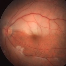
 Berlin's Edema
Apr 8 2019 by Gary R. Cook, MD, FACS
39-year-old white female with geographic area of retinal whitening ( Berlin's edema) without hemorrhage in the midperiphery secondary to blunt trauma; V.A. = 20/25
Imaging device: Topcon VT-50
Condition/keywords: Berlin's edema, blunt trauma, retinal edema
-
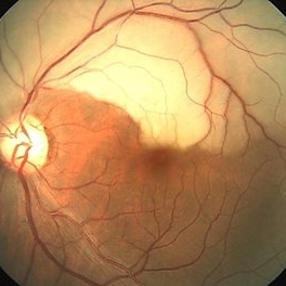
 Branch Retinal Artery Occlusion
Dec 19 2025 by Gayathri M S
Multicolor Reflectance and Blue Reflectance of a 55 yr male patient with blurring since 1 month shows classical sectoral retinal whitening.
Photographer: Gayathri MS
Imaging device: Heidelberg Spectralis
Condition/keywords: blue reflectance, branch retinal artery occlusion (BRAO), multicolor
-
 Branch Retinal Artery Occlusion With Calcium Embolus at the Disc - Fundus Photo
Apr 7 2018 by Rameez N Hussain, MD
Acute branch retinal artery occlusion with a calcium embolus at the disc with retinal whitening in the area of retinal edema.
Photographer: DR RAMEEZ N HUSSAIN
Imaging device: zeiss
Condition/keywords: branch retinal artery occlusion (BRAO), embolus, fundus photograph, retinal edema
 Loading…