Search results (46 results)
-
 Acute Idiopathic Occlusive Retinal Vasculitis
May 31 2014 by Hamid Ahmadieh, MD
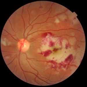
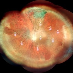
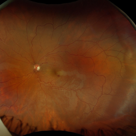
Color fundus photograph of the right eye of a 28-year-old woman with sudden drop of vision due to acute occlusive retinal vasculitis leading to extensive nerve fiber layer infarction and retinal hemorrhages.
Photographer: Naghmeh Nozhat, Negah Eye Center, Tehran
Condition/keywords: color fundus photograph, cotton wool spots, retinal hemorrhage, retinal ischemia
-
 Acute Idiopathic Occlusive Retinal Vasculitis
May 31 2014 by Hamid Ahmadieh, MD
Color fundus photograph of the left eye of a 28-year-old woman with acute drop of vision due to occlusive retinal vasculitis leading to extensive nerve fiber layer infarction and retinal hemorrhages.
Photographer: Naghmeh Nozhat, Negah Eye Center, Tehran
Condition/keywords: color fundus photograph, cotton wool spots, retinal hemorrhage, retinal ischemia
-
 Central Retinal Artery Occlusion
May 16 2017 by Olivia Rainey
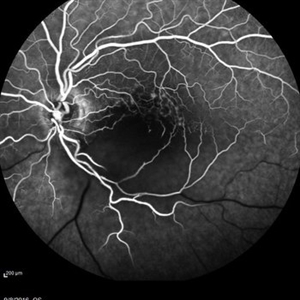
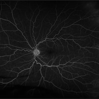
Fluorescein angiogram of an 66-year-old female with a central retinal artery occlusion affecting her left eye.
Photographer: Olivia Rainey
Imaging device: Heidelberg Spectralis
Condition/keywords: 50 degrees, central retinal artery occlusion (CRAO), fluorescein angiogram (FA), left eye, mid phase, retinal ischemia
-
 Central Retinal Artery Occlusion With Cilioretinal Sparing
Apr 4 2018 by Soumya Venkatesh
Fundus photograph of a 23-year-old gentleman presenting with sudden loss of vision 2 days prior to presentation. He underwent all relevant investigations and found to have APLA positive. He also had dengue serology positive. On follow up, his retinal edema reduced unmasking the underlying hemorrhages( flame shaped).
Photographer: Soumya Harapanahalli Venkatesh, JSS university, Karnataka, India
Condition/keywords: central retinal artery occlusion (CRAO), cherry red spot, cilioretinal sparing, retinal ischemia
-
 Central Retinal Vein Occlusion with Retinal Neovascularization
Jan 19 2022 by Olivia Rainey
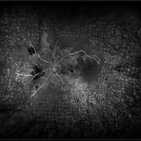
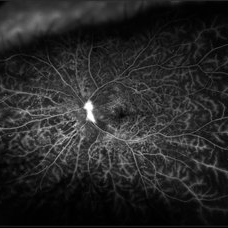
Ultra-widefield fluorescein angiogram of a 56-year-old male with a Central Retinal Vein Occlusion with Retinal Neovascularization affecting his left eye. The patient presented on 1/19/2022 with scNLP vision in the left eye. The patient has good PRP, however areas of ischemia still remain untreated by laser. He also has severe neovascular glaucoma contributing to his poor vision.
Photographer: Olivia Rainey, OCT-C, COA
Imaging device: Optos California
Condition/keywords: central retinal vein occlusion (CRVO), FA early phase, fluorescein angiogram (FA), hemorrhage, ischemic CRVO, left eye, neovascular glaucoma, Optos, pan-retinal photocoagulation (PRP), retinal ischemia, retinal neovascularization, ultra-wide field imaging
-
 Central Retinal Vein Occlusion with Severe Retinal Ischemia
Jan 19 2022 by Olivia Rainey
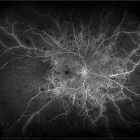
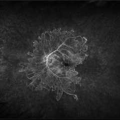
Ultra-widefield fluorescein angiogram of a 56-year-old male with a Central Retinal Vein Occlusion with Severe Retinal Ischemia affecting his right eye. The patient presented on 1/19/2022, sc20/20-2 vision in the right eye. The patient has had a good response to Eylea with complete resolution of edema. The physician is considering PRP to ischemic periphery in the future and given the degree of ischemia in both eyes, she recommends that the patient's PCP check carotid Doppler US.
Photographer: Olivia Rainey, OCT-C, COA
Imaging device: Optos California
Condition/keywords: central retinal vein occlusion (CRVO), FA late phase, fluorescein angiogram (FA), ischemic CRVO, Optos, retinal ischemia, ultra-wide field imaging
-
 Diabetic Retinopathy
Oct 2 2013 by Jerald A. Bovino, MD
This patient with long standing diabetes has peripheral non-perfusion.
Condition/keywords: retinal ischemia
-
 Familial Exudative Vitreo-retinopathy
Jul 6 2021 by Akansha Sharma
Color photo montage of 21-year-old male with familial exudative vitreoretinopathy in an amblyopic eye.
Photographer: Dr. Akansha Sharma-Retina Foundation, Ahmedabad
Condition/keywords: familial exudative vitreoretinopathy (FEVR), retinal ischemia
-
 Familial Exudative Vitreo-retinopathy
Jul 6 2021 by Akansha Sharma
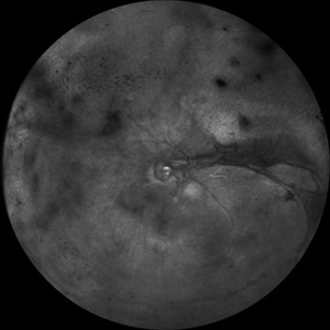
Infra-red capture of 5-year-old male with familial exudative vitro-retinopathy with disc pallor.
Photographer: Dr. Akansha Sharma-Retina Foundation, Ahmedabad
Condition/keywords: familial exudative vitreoretinopathy (FEVR), retinal ischemia
-
 Hypertensive Retinopathy
Aug 24 2012 by Geoffrey G. Emerson, MD, PhD, FASRS
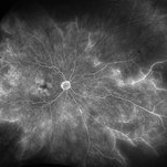
A 35-year-old man has headaches and decreased vision. The right eye measures 20/25 and the left eye measures 3/200. The blood pressure measures 180/110. This fluorescein angiogram shows dilated capillaries and capillary dropout in the central macula of the left eye.
Photographer: Geoffrey Emerson, MD, PhD, Retina Center, Minneapolis
Condition/keywords: hypertensive retinopathy, papilledema, retinal ischemia
-
 Ischemic Proliferative Diabetic Retinopathy
Aug 22 2012 by Edwin H. Ryan, MD
Fundus photograph of 35-year-old poorly-controlled diabetic woman, 6/200 vision.
Photographer: Edwin Ryan Jr. MD, VitreoRetinal Surgery, PA
Condition/keywords: retinal ischemia
-
 Ischemic Proliferative Diabetic retinopathy
Aug 22 2012 by Edwin H. Ryan, MD
Fluorescein angiogram of 35-year-old poorly-controlled diabetic woman, 6/200 vision.
Photographer: Edwin Ryan Jr. MD, VitreoRetinal Surgery, PA
Condition/keywords: retinal ischemia
-
---thumb.jpg/image-square;max$300,300.ImageHandler) Lupus Vasculitis Angiogram
Lupus Vasculitis Angiogram
Feb 13 2013 by From the Collections of Thomas M. Aaberg, MD and Thomas M. Aaberg Jr., MD
FA, lupus vasculitis angiogram.
Condition/keywords: lupus, obliterative peripheral vasculitis, retinal ischemia
-
 Marked Retinal Ischemia in Patient with Mixed Connective Tissue Disease
Feb 26 2013 by Sharon Fekrat, MD FACS FASRS
Fluorescein angiogram of the right eye of a 27-year-old female with mixed connective tissue disease and marked retinal ischemia. Panretinal laser photocoagulation (PRP) has been performed for neovascularization elsewhere (NVE).
Condition/keywords: mixed connective tissue disease, retinal ischemia
-
 Moderate Nonproliferative Diabetic Retinopathy
Mar 13 2025 by Drew Mitchell
Fluorescein angiography on a patient with Moderate Nonproliferative Diabetic Retinopathy at 5 minutes.
Photographer: Drew Mitchell OCT-C
Imaging device: Optos California
Condition/keywords: Diabetes, nonproliferative diabetic retinopathy, OPTOS CALIFORNIA, retinal ischemia
-
 Ocular Ischemic Syndrome/ Severe NPDR
Oct 6 2021 by Becca Harris
53 year old female with Severe NPDR and Ocular Ischemic Syndrome.
Photographer: Becca Harris
Imaging device: Optos California
Condition/keywords: Diabetic Retinopathy, left eye, nonproliferative diabetic retinopathy, ocular ischemic syndrome, optos, retinal ischemia
-
 Paracentral Acute Middle Maculopathy
Oct 22 2019 by Jeffrey G. Gross, MD, FASRS
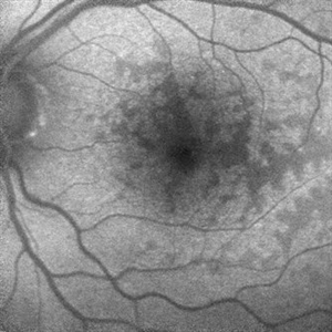
Fundus autofluorescence photograph of 75-year-old white male with 6 day history of acute vision loss. 20/40
Photographer: Tammy McLaughlin
Imaging device: Heidelberg Spectralis
Condition/keywords: paracentral acute middle maculopathy, retinal ischemia
-
 Paracentral Acute Middle Maculopathy
Oct 22 2019 by Jeffrey G. Gross, MD, FASRS
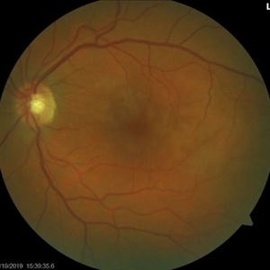
Color photograph of 75-year-old white male with 6 day history of acute vision loss. 20/40
Photographer: Tammy McLaughlin
Imaging device: Zeiss Visucam
Condition/keywords: paracentral acute middle maculopathy, retinal ischemia
-
 Paracentral Acute Middle Maculopathy
Oct 22 2019 by Jeffrey G. Gross, MD, FASRS
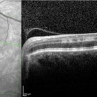
OCT of 75-year-old white male with 6 day history of acute vision loss. 20/40
Photographer: Tammy McLaughlin
Imaging device: Heidelberg Spectralis
Condition/keywords: paracentral acute middle maculopathy, retinal ischemia
-
 Paracentral Acute Middle Maculopathy
Oct 22 2019 by Jeffrey G. Gross, MD, FASRS
FA of 75-year-old white male with 6 day history of acute vision loss. 20/40
Photographer: Tammy Mclaughlin
Imaging device: Zeiss Visucam
Condition/keywords: FA late phase, paracentral acute middle maculopathy, retinal ischemia
-
 Paracentral Acute Middle Maculopathy (PAMM)
Oct 22 2019 by Jeffrey G. Gross, MD, FASRS
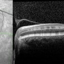
OCT of 75-year-old white male with 6 day history of acute vision loss. 20/40
Photographer: Tammy McLaughlin
Imaging device: Heidelberg Spectralis
Condition/keywords: paracentral acute middle maculopathy, retinal ischemia
-
 Patchy Ischemic Whitening in Sturge Weber Syndrome
Nov 18 2024 by Edward F. Hall, MD, FASRS
Left fundus photograph of a 44-year-old man showing patchy ischemic retinal whitening associated with Sturge-Weber Syndrome. The precise cause of this rare complication remains unclear, but it may be linked to choroidal vascular congestion and a compartment syndrome-like effect on the local retinal arteriolar circulation. OCT imaging confirmed inner retinal ischemia and thickening
Photographer: Karissa Kuhl
Imaging device: Optos
Condition/keywords: retinal ischemia, Sturge Weber Syndrome
-
 Peripheral Retinal Ischemia
Apr 26 2018 by Olivia Rainey
Ultra-wide field fluorescein angiogram of a 55-year-old female with peripheral retinal ischemia affecting her left eye. CTA head and neck performed on 11/16/15 and showed calcified atherosclerotic plaque involving the intracranial internal carotid arteries with resulting luminal narrowing. Intracranial vertebral arteries have smooth luminal contours. CTA neck normal. Likely from internal carotid plaques. Sickle cell disease came back negative.
Photographer: Olivia Rainey
Imaging device: Optos California
Condition/keywords: fluorescein angiogram (FA), fluorescein leakage, left eye, Optos, retinal ischemia, ultra-wide field imaging
-
 Proliferative Diabetic Retinopathy and SC Disease
Aug 27 2021 by Caesar K. Luo, MD, FASRS
53 year-old male with SC disease complicated by proliferative diabetic retinopathy with severe peripheral non perfusion and small, central retained island.
Photographer: Fred Hanamoto, Bay Area Retina Associates
Imaging device: Optos California
Condition/keywords: capillary nonperfusion, peripheral ischemia, proliferative diabetic retinopathy (PDR), retinal ischemia, sickle cell retinopathy
-
 Proliferative Diabetic Retinopathy with Retinal Ischemia
Mar 29 2019 by Olivia Rainey
Ultra-wide field fluorescrein angiogram of a 42-year-old female with proliferative diabetic retinopathy with retinal ischemia affecting her left. Patient had been noticing a vision decline and floaters over the past few months. She was treated with Avastin in her right eye and then her left one week later.
Photographer: Olivia Rainey
Imaging device: Optos
Condition/keywords: diabetes, diabetic macular edema, early phase, fluorescein angiogram (FA), fluorescein leakage, neovascularization of the disc (NVD), Optos, proliferative diabetic retinopathy (PDR), retinal ischemia, ultra-wide field imaging
 Loading…