Search results (648 results)
-
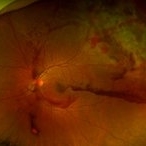
 5 Watt Blue Laser Pointer, Retinal Hemorrhage
Aug 30 2018 by John S. King, MD
5 watt laser pointer (class 4 laser pointer that can burn skin and material) to eye caused this hemorrhage as a result of injury to the retinal venule (see photo). Seen by Dr. Arnold, who sent this patient to Dr. Ware. Fortunately, fovea spared.
Photographer: Stacy
Imaging device: Topcon
Condition/keywords: laser pointer maculopathy, retinal hemorrhage
-
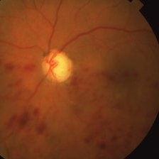
 5 Watt Blue Laser Pointer, Retinal Hemorrhage
Aug 30 2018 by John S. King, MD
5 watt laser pointer (class 4 laser pointer that can burn skin and material) to eye caused this hemorrhage as a result of injury to the retinal venule (see photo). Seen by Dr. Arnold, who sent this patient to Dr. Ware. Fortunately, fovea spared.
Imaging device: Optos
Condition/keywords: laser pointer retinopathy, retinal hemorrhage
-
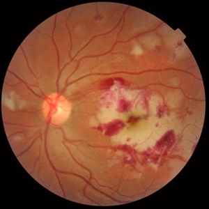
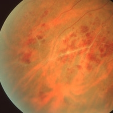
 Acute Idiopathic Occlusive Retinal Vasculitis
May 31 2014 by Hamid Ahmadieh, MD
Color fundus photograph of the right eye of a 28-year-old woman with sudden drop of vision due to acute occlusive retinal vasculitis leading to extensive nerve fiber layer infarction and retinal hemorrhages.
Photographer: Naghmeh Nozhat, Negah Eye Center, Tehran
Condition/keywords: color fundus photograph, cotton wool spots, retinal hemorrhage, retinal ischemia
-
 Acute Idiopathic Occlusive Retinal Vasculitis
May 31 2014 by Hamid Ahmadieh, MD
Color fundus photograph of the left eye of a 28-year-old woman with acute drop of vision due to occlusive retinal vasculitis leading to extensive nerve fiber layer infarction and retinal hemorrhages.
Photographer: Naghmeh Nozhat, Negah Eye Center, Tehran
Condition/keywords: color fundus photograph, cotton wool spots, retinal hemorrhage, retinal ischemia
-
 Anemic Retinopathy in a Young Female
Jan 15 2022 by KRISHNENDU NANDI, MS
Fundus photograph of a 29-year-old female presented with retinal hemorrhages in both eyes with decrease in vision for 1 month. Hemoglobin level was 5.9gm/dl, suggestive of anemic retinopathy in both eyes.
Photographer: Krishnendu Nandi, Netralayam Eye Care Centre, Kolkata, India
Imaging device: Topcon
Condition/keywords: anaemic retinopathy, anemic retinopathy, retinal hemorrhage
-
 Anemic Retinopathy Related Retinal Hemorrhages
Nov 5 2019 by Chinmayi Vyas
Anemic retinopathy related retinal hemorrhages in a 24 years old male with Hb of 4.2gm/ dl. The manifestations of anemic retinopathy are nonspecific and may closely simulate hypertensive or diabetic retina. Retinal changes in anemia are cotton wool spots, venous tortuosity, and hemorrhages which may be present at all levels of the retina and choroid. All retinal hemorrhages can occur when Hb falls below 8 g/100 ml or if the platelet count falls below 50,000/cumm. The combination of severe anemia and thrombocytopenia is likely to produce retinal hemorrhages. The Roth’s spots or white centre hemorrhages are typically associated with bacterial endocarditis , anemia and other systemic conditions. The white center is suspected to represents focal ischemia, inflammatory or infectious infiltrate, fibrin or accumulation of neoplasticism cells.
Photographer: Dr Chinmayi Vyas
Condition/keywords: retinal hemorrhage
-
 Best Disease
Apr 8 2019 by Gary R. Cook, MD, FACS
Left eye of the patient with Best disease and an active CNV OD showing resolving hemorrhage from prior CNV OS.
Condition/keywords: Best disease, retinal hemorrhage, vitelliform macular dystrophy
-
 Blunt Ocular Trauma with Commotio Retinae
Nov 5 2019 by Nichole Lewis
11-year-old male with blunt ocular trauma from a soccer ball. Commotio Retinae, retinal hemorrhages, vitreous hemorrhage, multiple retinal tears and a traumatic macular hole. VA 20/70.
Photographer: Nichole Lewis
Imaging device: Optos
Condition/keywords: blunt trauma, commotio retinae, retinal hemorrhage, retinal tear, traumatic macular hole, vitreous hemorrhage
-
 BRVO With Macular Involvement
Feb 19 2015 by H. Michael Lambert, MD
BRVO, superotemporal, macular edema.
Condition/keywords: branch retinal vein occlusion (BRVO), macular edema, retinal hemorrhage
-
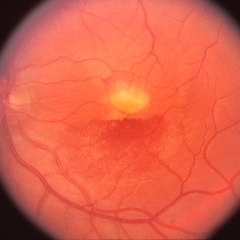
 Central Retinal Vein Occlusion
Feb 20 2013 by From the Collections of Thomas M. Aaberg, MD and Thomas M. Aaberg Jr., MD
Retinal hemorrhages Central Retinal Vein Occlusion Fundus Photograph
Condition/keywords: central retinal vein occlusion (CRVO), retinal hemorrhage
-
 Choriodal Rupture 004 - Fundus Autoflurescence - 6 Month Follow Up
Mar 11 2013 by Suber S. Huang, MD, MBA, FASRS
40-year-old male sustained blunt trauma OD with orbital fracture and choroidal rupture subjacent to inferior arcade with blood, subretinal fluid, and exudate extending to fovea with CF 2 feet at presentation 9-10-12. On follow up 3-11-12, vision improved to 20/400 with resolutionof hemorrhage, normal OCT, but speckling of foveal RPE and PMB consistent with damage.
Photographer: Mark Harrod
Condition/keywords: autofluorescence imaging, blunt trauma, choroidal hemorrhage, choroidal rupture, orbital fracture, retinal hemorrhage, submacular hemorrhage
-
 Choriodal Rupture - 003 - 6 Month Follow Up
Mar 11 2013 by Suber S. Huang, MD, MBA, FASRS
40 -year-old male sustained blunt trauma OD with orbital fracture and choroidal rupture subjacent to inferior arcade with blood, subretinal fluid, and exudate extending to fovea with CF 2 feet at presentation 9-10-12. On followup 3-11-12, vision improved to 20/400 with resolutionof hemorrhage, normal OCT, but speckling of foveal RPE and PMB consistent with damage.
Photographer: Mark Harrod
Condition/keywords: autofluorescence imaging, blunt trauma, choroidal hemorrhage, choroidal rupture, orbital fracture, retinal hemorrhage, submacular hemorrhage
-
 Choriodal Rupture - AW 001 - Initial Presentation
Mar 11 2013 by Suber S. Huang, MD, MBA, FASRS
40-year-old male sustained blunt trauma OD with orbital fracture and choroidal rupture subjacent to inferior arcade with blood, subretinal fluid, and exudate extending to fovea with CF 2 feet at presentation 9-10-12. On followup 3-11-12, vision improved to 20/400 with resolution of hemorrhage, normal OCT, but speckling of foveal RPE and PMB consistent with damage.
Photographer: Mark Harrod
Condition/keywords: autofluorescence imaging, blunt trauma, choroidal hemorrhage, choroidal rupture, orbital fracture, retinal hemorrhage, submacular hemorrhage
-
 Choriodal Rupture - AW 002 - 6 Month F/U
Mar 11 2013 by Suber S. Huang, MD, MBA, FASRS
40-year-old male sustained blunt trauma OD with orbital fracture and choroidal rupture subjacent to inferior arcade with blood, subretinal fluid, and exudate extending to fovea with CF 2 feet at presentation 9-10-12. On follow up 3-11-12, vision improved to 20/400 with resolutionof hemorrhage, normal OCT, but speckling of foveal RPE and PMB consistent with damage.
Photographer: Mark Harrod
Condition/keywords: autofluorescence imaging, blunt trauma, choroidal hemorrhage, choroidal rupture, orbital fracture, retinal hemorrhage, submacular hemorrhage
-
 CNVM due to Pathologic Myopia
Apr 2 2019 by Gary R. Cook, MD, FACS
55-year-old Asian male with -9.50D myopia with a visible (Type I) CNVM and thin hemorrhage in the macula: V.A.= 20/200
Imaging device: Topcon VT-50
Condition/keywords: choroidal neovascular membrane (CNVM), high myopia, pathologic myopia, retinal hemorrhage
-
 CNVM Due to Pathologic Myopia
Apr 2 2019 by Gary R. Cook, MD, FACS
Fluorescein angiogram frame of the left eye of the 55-year-old Asian male with -9.50D myopia OS with a subfoveal CNVM and hemorrhage secondary to high myopia; V.A.= 20/200.
Imaging device: Topcon VT-50
Condition/keywords: choroidal neovascular membrane (CNVM), high myopia, pathologic myopia, retinal hemorrhage, subfoveal choroidal neovascularization
-
 Commotio Retinae with Hemorrhage
Mar 27 2018 by Nichole Lewis
Commotio Retinae with hemorrhage.
Photographer: Nichole Lewis
Condition/keywords: commotio retinae, retinal hemorrhage
-
 Commotio Retinae with Retinal Hemorrhages
Mar 27 2018 by Nichole Lewis
14-year-old male hit in the right eye with a stick. Commotio Retinae with retinal hemorrhages and peripapillary hemorrhage.
Photographer: Nichole Lewis
Condition/keywords: commotio retinae, peripapillary hemorrhage, retinal hemorrhage
-
---thumb.JPG/image-square;max$300,300.ImageHandler) CRVO
CRVO
Oct 27 2012 by Mallika Goyal, MD
Fundus photograph of a 52-year-old gentleman with fresh CRVO; retinal haemorrhages all quadrants with optic disc edema and macular edema.
Condition/keywords: central retinal vein occlusion (CRVO), retinal hemorrhage
-
 Dengue Retinitis
Oct 27 2012 by Mallika Goyal, MD
Right eye of a 37-year old non-diabetic, non-hypertensive lady recovering from dengue fever with bilateral dengue retinitis shows vitreous haze, retinal hemorrhages and exudates over the posterior pole. Subsequently she had dengue foveolitis, NVD and vitreous haemorrhage in this eye.
Photographer: Mallika Goyal, MD
Condition/keywords: Dengue retinitis, retinal hemorrhage, vitreous haze
-
 Dengue Retinitis
Oct 27 2012 by Mallika Goyal, MD
Left eye of a 37-year-old lady recovering from dengue fever with bilateral dengue retinitis shows retinal hemorrhages and exudates over the posterior pole.
Photographer: Mallika Goyal, MD
Condition/keywords: bilateral dengue retinitis, exudates over the posterior pole, retinal hemorrhage
-
 Eales Disease
Apr 1 2019 by Gary R. Cook, MD, FACS
25-year-old Vietnamese male with peripheral retinal vasculitis, vaso-occlusion, and retinal hemorrhages secondary to Eales Disease; V.A.= 20/20.
Imaging device: Topcon VT-50
Condition/keywords: Eales disease, retinal hemorrhage, vaso-occlusive disease, vasoocclusive retinopathy
-
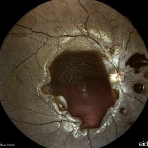
 Gross Pathology - Retinal Detachment and Retinal Hemorrhage
Feb 20 2013 by From the Collections of Thomas M. Aaberg, MD and Thomas M. Aaberg Jr., MD
Gross specimen Retinal Detachment Retinal Hemorrhage
Condition/keywords: gross specimen, retinal hemorrhage
-
 Hemi-CRVO
Mar 27 2019 by Gary R. Cook, MD, FACS
78-year-old African American female patient with COAG and ischemic inferior hemi-CRVO OS; V.A. = HM 1 ft.
Imaging device: Topcon VT-50
Condition/keywords: chronic open-angle glaucoma (COAG), hemi CRVO, retinal hemorrhage
-
---thumb.JPG/image-square;max$300,300.ImageHandler) hypertensive retinopathy
hypertensive retinopathy
Nov 3 2012 by Mallika Goyal, MD
Right eye of a 30-year-old hypertensive gentleman shows hypertensive retinopathy grade 4 with retinal hemorrhages, exudates and disc edema.
Photographer: Mallika Goyal, MD
Condition/keywords: disc edema, hypertensive retinopathy, retinal hemorrhage
 Loading…