Search results (47 results)
-
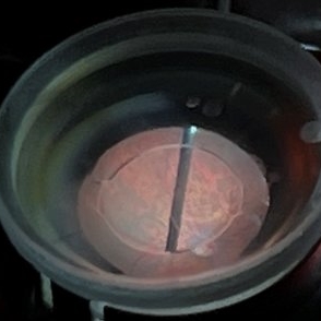
25 Gauge Vitrectomy Membrane Shaving
Jan 31 2015 by Thomas A. Ciulla, MD, MBA, FASRS
Membrane shaving of dense membranes in diabetic traction detachment using 25 gauge vitrectomy.
Condition/keywords: diabetes, pars plana vitrectomy (PPV), retina surgery, tractional retinal detachment, vitreoretinal surgery
-
 Brilliant Blue Dye Injected in a Case of Macular Hole to Stain the ILM
Feb 4 2022 by Manish Nagpal, MD, FRCS (UK), FASRS
Intraoperative still of a brilliant blue dye being injected to stain the ILM.
Photographer: Manish Nagpal, Director, Retina Foundation, Ahmedabad
Imaging device: Sony PMW -10 MD surgical camera
Condition/keywords: brilliant blue, ILM flap, ILM staining, macular hole, retina, retina surgery
-
 Dexamethasone Implant
Jul 3 2021 by Gerardo Rivera Arroyo
42-year-old male, operated on for vitrectomy plus scleral buckling plus silicone plus dexamethasone implant for inferior retinal detachment with PVR.
Photographer: Rosa Elizabeth Moreno Anda, MD, Hospital Central Militar CDMX
Condition/keywords: dexamethasone implant, retina surgery, vitrectomy
-
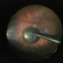
 Direct lens, indirect vision.
May 19 2022 by ALLAN GOMES DA SILVA
External view of vitrectomy under direct macular lens.
Photographer: Allan Gomes da Silva
Imaging device: Wide-angle camera - 26mm; f1.5; ISO 64; 208mm; 1/120 sec
Condition/keywords: retina surgery, vitreomacular surgery
-
 ILM Peeling in Progress
Feb 4 2022 by Manish Nagpal, MD, FRCS (UK), FASRS
Intraoperative shot of ILM peeling in progress using forceps.
Photographer: Manish Nagpal, Director, Retina Foundation, Ahmedabad
Imaging device: Sony PMW -10 MD surgical camera
Condition/keywords: ILM flap, ILM staining, internal limiting membrane (ILM) peeling, macular hole, retina, retina surgery
-
 Neovascular Glaucoma
Jan 3 2020 by Manuel Ángel Alcántara Delgado, MD
Slit lamp photograph of a 65-year-old man with diabetic retinopathy and previous history of phaco-vitrectomy.
Photographer: Manuel Ángel Alcántara Delgado, CMN SXXI, Mexico City
Condition/keywords: diabetes, diabetic retinopathy, neovascular glaucoma, neovascularization (NV), retina surgery, retina surgery complications
-
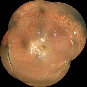
 Optos Silverstone Fundus Image of a 4-Point Scleral Fixation Akreos AO60 with Gore Tex Suture
Dec 5 2021 by Jesus Lozano, MD
Optos Silverstone fundus image of a 54-year-old man, 6 months after 4-point scleral fixation Akreos AO60 with Gore Tex suture plus PPV who had a severe traumatic iris defect and was aphakic after ocular trauma.
Photographer: Yair Bet Yosef, Hadassah Medical Center. Israel
Imaging device: Optos Silverstone fundus image
Condition/keywords: fundus photograph, Gore Tex Suture, macula, ocular trauma, retina surgery, scleral fixation
-
 Perforating Ocular Injury
Apr 18 2022 by Franco Benvenuto, MD
16 Y/O Male with history of perforating injury with a metal nail, days after nail extraction laser photocoagulation around the exit injury was performed.
Photographer: Franco Benvenuto, Universidad de Buenos Aires, Argentina
Condition/keywords: laser photocoagulation, retina surgery, Trauma
-
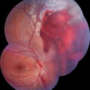
 Persistent Fetal Vasculature Before and After Surgery
Jan 13 2020 by Sophia El Hamichi, MD
A: Fundus photograph of a 15-month-old male showing a persistent fetal vasculature with secondary tractional detachment B: Fundus photograph of the patient eye after repair surgery with flat retina
Photographer: Abby Orcutt-Hayes, Murray Ocular Oncology and Retina
Condition/keywords: persistent fetal vasculature (PFV), persistent hyperplastic primary vitreous (PHPV), retina surgery, tractional retinal detachment
-
 PPV retained cataract
Apr 19 2023 by Denica Rodriguez
A 46-year-old male with hypermature dense cataract. Patient got a piece of metal in his eye when he was 5 years old and was not able to see since. Patient was having cataract surgery and phacodonesis was present. The lens dropped to the back of the eye. Patient had to have another surgery to do vitrectomy. The lens removal was done with a fragmatome handpiece.
Photographer: Denica Rodriguez COA, ST
Imaging device: Zeiss Microscope with resight
Condition/keywords: cataract, dropped nucleus, fragmatome, lens capsule, ocular trauma, pars plana vitrectomy (PPV), retained lens fragments, Retina, retina surgery, traumatic cataract
-
---thumb.jpg/image-square;max$300,300.ImageHandler) Proliferative Vitreoretinopathy (PVR)
Proliferative Vitreoretinopathy (PVR)
Feb 13 2013 by From the Collections of Thomas M. Aaberg, MD and Thomas M. Aaberg Jr., MD
Recurrent retinal detachment, PVR.
Condition/keywords: proliferative vitreoretinopathy (PVR), retina surgery
-
 Recurrent Retinal Detachment with PVR with Subretinal Oil after Retinal Detachment with Silicone Oil
Feb 2 2022 by Manish Nagpal, MD, FRCS (UK), FASRS
Intraoperative photo of inferior retinal contraction due to PVR and presence of subretinal silicone oil globule noted.
Photographer: Manish Nagpal, Retina Foundation, Ahmedabad, Gujarat
Imaging device: Sony PMW -10 MD surgical camera
Condition/keywords: proliferative vitreoretinopathy (PVR), retina, retina surgery, retina surgery complications, silicone oil, subretinal
-
 Retinal Detachment
Feb 4 2022 by Manish Nagpal, MD, FRCS (UK), FASRS
Intraoperative still of a retinal detachment with macula involvement.
Photographer: Manish Nagpal, Director, Retina Foundation, Ahmedabad
Imaging device: Sony PMW -10 MD surgical camera
Condition/keywords: retina, retina surgery
-
 Retinal Detachment
Feb 4 2022 by Manish Nagpal, MD, FRCS (UK), FASRS
Intraoperative still of a retinal detachment undergoing vitrectomy.
Photographer: Manish Nagpal, Director, Retina Foundation, Ahmedabad
Imaging device: Sony PMW -10 MD surgical camera
Condition/keywords: retina, retina surgery, Retinal Detachment
-
 Retinal Detachment with Early PVR Changes Including Star Folds
Feb 4 2022 by Manish Nagpal, MD, FRCS (UK), FASRS
Intraoperative still photo of a retinal detachment and PVR with star folds undergoing vitrectomy.
Photographer: Manish Nagpal, Director, Retina Foundation, Ahmedabad
Imaging device: Sony PMW -10 MD surgical camera
Condition/keywords: proliferative vitreoretinopathy (PVR), retina, retina surgery, Retinal Detachment, star folds
-
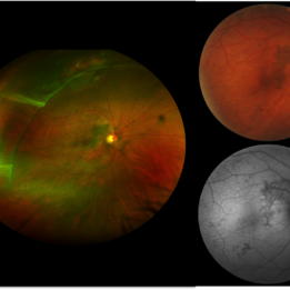 Retinal Folds After Surgery
Jun 23 2016 by Andrea Arriola-Lopez, MD MSc
45-year-old man with history of rhegmatogenous retinal detachment and segmental scleral buckle from MIX to MXII, SF6 and cryotherapy on right eye was performed. Radial folds on indentation was seen after surgery. Three weeks later, inferior macular folds was found. The patient was asymptomatic. Observation was decided. Retina remains attach. On top, close up to macular area shows inferior folds far from fovea. Bottom, red free photograph shows no RPE changes on the same retina fold area.
Photographer: Andrea E. Arriola-López MD MSc
Imaging device: OPTOS
Condition/keywords: macular fold, retina surgery, scleral buckle
-
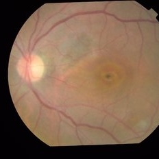 Retinography
May 24 2022 by ALLAN GOMES DA SILVA
Macular pigmentation? Closed macular hole appearance after pars plana vitrectomy, enlarged ILM peeling, approximation of hole edges and foveal repositioning maneuver.
Condition/keywords: macular hole, retina surgery
-
 Rhegmatogenous retinal detachment in a 27 year old man.
Mar 14 2022 by Jesus Lozano, MD
27 year old man with a rhegmatogenous retinal detachment macula off.
Photographer: Dr. Avi Schwalb, Hillel Yaffe Medical Center, Israel.
Imaging device: Optos Silverstone
Condition/keywords: retina surgery, retinal detachment with retinal defect, scleral band, scleral buckle
-
 Scleral buckle indent s/p retina surgery
Dec 13 2023 by rahul saradge
Scleral buckle indent s/p retina surgery
Photographer: Saloni Mishra , Isha Netralaya.
Imaging device: optos
Condition/keywords: Optos, Retina buckle, retina surgery, scleral buckle, ultra-wide field imaging
-
 Scleral Buckle Surgery
Mar 14 2022 by Jesus Lozano, MD
27 year old man after 1 Week Scleral Buckle Surgery. 4.0mm Silicon Strip. Retina attached.
Photographer: Dr. Avi Schwalb, Hillel Yaffe Medical Center, Israel.
Imaging device: Optos Silverstone
Condition/keywords: retina surgery, retinal detachment with retinal defect, scleral band, scleral buckle
-
 Shafer's Sign
Jan 3 2020 by Manuel Ángel Alcántara Delgado, MD
Slit lamp photograph of a 58-year-old man with rhegmatogenous retinal detachment and tobacco dust presence.
Photographer: Manuel Ángel Alcántara Delgado, CMN SXXI, Mexico City
Condition/keywords: acute retinal detachment, retina surgery, vitrectomy
-
 Silicon Oil in CT brain
Aug 5 2022 by Jesus Lozano, MD
An 65-year-old woman was taken to the Emergency Department after a fall. CT brain imaging demonstrated a well-defined, homogenous, hyperdense mass in the posterior segment of the right eye. Detailed history revealed previous vitreoretinal procedures for multiple retinal detachments. Ophthalmological examination confirmed presence of silicone oil in this eye.
Photographer: Dr. Jesus Lozano Gutierrez
Condition/keywords: retina surgery
-
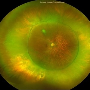
 SILICONE OIL FILLED VITRECTOMISED EYE STATUS POST PARS PLANA VITRECTOMY WITH SILICONE OIL INFUSION IN A CASE OF RHEGMATOGENOUS RETINAL DETACHMENT
Oct 19 2022 by Akansha Sharma
COLOUR FUNDUS MONTAGE OF A 11 YEAR OLD MALE WITH SILICONE OIL FILLED VITRECTOMISED EYE STATUS POST PARS PLANA VITRECTOMY WITH SILICONE OIL INFUSION IN A CASE OF RHEGMATOGENOUS RETINAL DETACHMENT
Photographer: Dr. Akansha Sharma-Retina Foundation, Ahmedabad
Condition/keywords: retina surgery, rhegmatogenous retinal detachment, silicone oil
-
 Spontaneously Dropped Lens in a Congenital Rubella Syndrome
Apr 30 2022 by NEIFFER RABELO
Intraoperative photograph of a 68-year-old patient with congenital rubella syndrome and light perception visual acuity since childhood. The image shows a pigmentary retinopathy and the lens spontaneously displaced into the vitreous cavity. The patient sought care complaining of a total and sporadic loss of vision that was hindering her in daily tasks. Surgery was indicated to remove the lens.
Photographer: Rodrigo Dos Anjos Versiani - Retina Institute - Belo Horizonte - Brazil
Imaging device: ZEISS OPMI LUMERA 700
Condition/keywords: dropped nucleus, retina surgery, rubella retinopathy
-
 Spontaneously dropped Nucleus In a Congenital Rubella Retina disease
Apr 29 2022 by NEIFFER RABELO
Intraoperative Fundus Photograph of an 68 years old with past medical history of
Photographer: Rodrigo Dos Anjos Vesiani, Retina Institute - Belo Horizonte - Brazil
Imaging device: OPMI LUMERA® 700
Condition/keywords: abnormal retina, dropped nucleus, retina surgery, rubella
 Loading…