Search results (27 results)
-
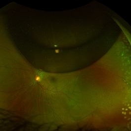
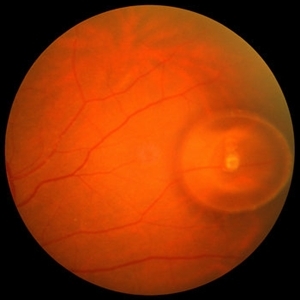
 C3F8 Gas Bubble for Retinal Tear/ Pneumatic Retinopexy
Jun 27 2013 by Jason S. Calhoun
Patient comes in with retinal detachment and a pneumatic retinopexy was performed. Gas bubble is visible with optic nerve reflected.
Photographer: Jason S. Calhoun, Mayo Clinic Jacksonville, Florida
Imaging device: TOPCON TRC 50-EX
Condition/keywords: pneumatic retinopexy
-
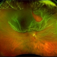
 Chronic Retinal Detachment after Pneumatic Retinopexy
Jan 8 2022 by Parnian Arjmand, MD, MSc, FRCSC, DABO
This is a fundus photo in the eye of a young phakic patent who presented with a 6 month history of "difficulty seeing at night" and subjective nasal "blurriness" in the left eye. There was a chronic temporal RD, OS, extending to the arcades (Mac on). This photo is week 1 s/p Pneumatic retinopexy with SF6 gas and laser retinopexy to temporal breaks (6 holes, lattice); no PVD. As you can see, there is a "bleb" of viscous schlieren given the chronic nature of this RD that persist posterior to the breaks and temporal to the macula. This type of sub retinal fluid may take months to years to resorb.
Condition/keywords: chronic retinal detachment, pneumatic retinopexy
-
---thumb.JPG/image-square;max$300,300.ImageHandler) Gas Bubble
Gas Bubble
Jul 12 2013 by Jason S. Calhoun
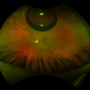
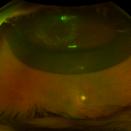
Patient had a pneumatic retinopexy for retinal detachment. Fundus shows C3F8 gas bubble superiorly.
Photographer: Jason S. Calhoun, Department of Ophthalmology, Mayo Clinic Jacksonville, Florida
Condition/keywords: pneumatic retinopexy
-
---thumb.JPG/image-square;max$300,300.ImageHandler) Gas Bubble
Gas Bubble
Jul 12 2013 by Jason S. Calhoun
Patient had a pneumatic retinopexy for retinal detachment. Fundus shows C3F8 gas bubble superiorly.
Photographer: Jason S. Calhoun, Department of Ophthalmology, Mayo Clinic Jacksonville, Florida
Condition/keywords: pneumatic retinopexy
-
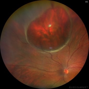 Giant Retinal Cyst
Sep 20 2025 by JORGE SOBERANES
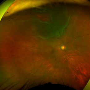
Fundus photograph of a 45-year-old-man with a large cyst on the nasal superior side of the retina. The patient had a history of a pneumatic retinopexy two years ago and the cyst has been there since that.
Photographer: Dr. Jorge Soberanes, Asociación para Evitar la Ceguera en México (APEC), UNAM
Condition/keywords: abnormal retina, pneumatic retinopexy, retinal cyst
-
 Giant Retinal Tear
Apr 29 2021 by Fong May Chew, FRCOphth, MBBS, BSc
Optos pictures of a 56-year-old man who presented with a giant retinal tear who extending from 7-12 o clock with a separate horseshoe tear at 1 o clock. Treated with pneumatic retinopexy.
Photographer: Hesham Hamli
Condition/keywords: giant retinal tear, pneumatic retinopexy
-
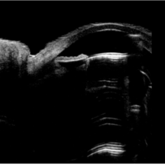 Intralenticular Gas
Aug 10 2024 by Varsha Reddy
Ultrasound biomicroscopy image of resolving intralenticular gas at office visit approximately 4 weeks later (B). The lens remained unchanged in thickness at 3.78 mm before and after resolution of the gas bubble, but developed a cataract.
Condition/keywords: intralenticular gas, pneumatic retinopexy, post-op
-
 Intralenticular Gas
Aug 10 2024 by Varsha Reddy
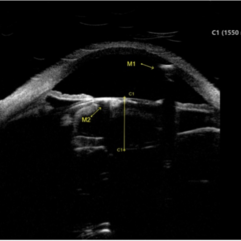
Ultrasound biomicroscopy image of gas in the anterior chamber (A, M1) and lenticular cortex (A, M2) of a 68-year-old man following pneumatic retinopexy. Patient presented with a macula-off retinal detachment requiring vitrectomy, with pneumatic retinopexy done in office at post-operative week 1 to supplement poor gas fill.
Condition/keywords: intralenticular gas, pneumatic retinopexy, post-op
-
 Pneumatic Retinopexy
Jan 9 2022 by Dante Akira Kondo Kuroiwa, MD, MBA
Superior rhegmatogenous retinal detachment with a horseshoe tear at 11 o'clock treated with pneumatic retinopexy.
Photographer: Dante Akira Kondo Kuroiwa, Federal University of Sao Paulo, Ophthalmology and Visual Sciences Department
Imaging device: Ultra-widefield Fundus Photography (Daytona-Optos)
Condition/keywords: pneumatic retinopexy
-
 Pneumatic Retinopexy
Jan 6 2025 by Mateus Queiroz Corrêa, MD
Fundus photograph of a pneumatic retinopexy. The upper photo taken just 30 minutes after C3F8 gas injection shows rhegmatogenous retinal detachment with superior temporal horseshoe tear and gas bubbles resembling fish-eggs. After two days with appropriated head position (botton photo), the retina is attached and laser photocoagulation was performed on the border of the break. A Single great gas bubble was formed.
Photographer: Mateus Queiroz Corrêa, Sorocada Eye Bank Hospital
Imaging device: Optos California
Condition/keywords: pneumatic retinopexy
-
 Pneumatic Retinopexy by Intravitreal Injection of Sulfur Hexafluoride Gas (SF6) at the Time of Diagnosis With Subsequent Application of 532 Nm Laser Around the Retinal Tear
Apr 4 2025 by Cesar Orlando Oviedo Vera
A 45-year-old male patient presented with a sudden onset of decreased visual acuity in the right eye, with a 24-hour progression. Upon examination, Image 1 revealed a superior rhegmatogenous retinal detachment in the right eye, with a retinal tear located between the 1 and 2 o'clock positions. Image 2: Pneumatic retinopexy by intravitreal injection of Sulfur Hexafluoride gas (SF6) at the time of diagnosis with subsequent application of 532 nm laser around the retinal tear.
Photographer: Cesar Orlando Oviedo Vera, Hospital Militar de Especialidades Oftalmológicas
Imaging device: Optos
Condition/keywords: Pneumatic Retinopexy, Retinal tear, Rhegmatogenous retinal detachment, SF6, Superior rhegmatogenous retinal detachment
-
 Sub Retinal Gas 1 Day Post Pneumatic Retinopexy
Jul 8 2016 by Asaf Friehmann
Fundus photograph of an 71-year-old male with a sub retinal C3F8 1 day after pneumatic retinopexy for the treatment of rhegmatogenous retinal detachment involving a single 1 hour horseshoe tear at 12 o'clock.
Photographer: Lilach Gorek
Condition/keywords: pneumatic retinopexy
-
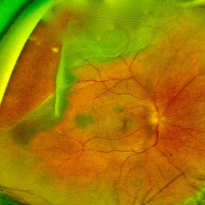
 Subretinal Gas After Pneumatic Retinopexy
Mar 6 2024 by James P Dossett, MD
Pseudocolor fundus photograph of a 68-year-old man who presented with a macula-on rhegmatogenous retinal detachment with a single horseshoe tear at 12 o'clock. Pneumatic retinopexy was performed with cryopexy to the retinal break. He returned to clinic three days later and the entire SF6 gas bubble was noted to have migrated to the subretinal space through the retinal break. Pars plana vitrectomy was performed that day with retinal reattachment and improvement in vision to 20/40 now 6 months postoperatively.
Imaging device: Optos
Condition/keywords: pneumatic retinopexy, subretinal gas bubble
-
 Superior Rhegmatogenous Retinal Detachment (RRD) in the Right Eye, With a Retinal Tear Located Between the 1 and 2 O'clock Positions
Apr 4 2025 by Cesar Orlando Oviedo Vera
A 45-year-old male patient presented with a sudden onset of decreased visual acuity in the right eye, with a 24-hour progression. Upon examination, Image 1 revealed a superior rhegmatogenous retinal detachment in the right eye, with a retinal tear located between the 1 and 2 o'clock positions. Image 2: Pneumatic retinopexy by intravitreal injection of Sulfur Hexafluoride gas (SF6) at the time of diagnosis with subsequent application of 532 nm laser around the retinal tear.
Photographer: Cesar Orlando Oviedo Vera, Hospital Militar de Especialidades Oftalmológicas
Imaging device: Optos
Condition/keywords: Pneumatic Retinopexy, Retinal tear, Rhegmatogenous retinal detachment, SF6, Superior rhegmatogenous retinal detachment
-
 Trapped Subretinal Gas Bubble
Oct 12 2012 by Jeffrey G. Gross, MD, FASRS
Trapped subretinal gas bubble after pneumatic retinopexy procedure.
Condition/keywords: pneumatic retinopexy, subretinal gas bubble
-
 Pneumatic Retinopexy Before & After
Apr 27 2023 by Jesus Lozano, MD
Pneumatic Retinopexy Before & After Optos Pictures. 47 year old man with a Superior Rhegmatogenous Retinal Detachment Macula Off (retinal tear at 01:00hrs) -1st Image: RRD macula off before treatment. -2nd Image: After 48 hrs from Pneumatic Retinopexy.
Imaging device: Optos
-
 Gardner Syndrome
Dec 12 2018 by John S. King, MD
66-year-old white male with Gardner Syndrome (colon resection in 1991), who has two children with Gardner Syndrome, presented to Dr. Zocchi with an RD in the fellow eye that was successfully repaired with a pneumatic retinopexy. Currently 20/20 OU with IOP of 7 OD and 14 OS; no RAPD; PCIOL OU. Dr. Zocchi got oral permission by the patient to have these put into the Retina Image Bank. Although the CHRPE like lesions (2 OD) are not bilateral, we both think these lesions represent "retinal pigment epithelial hamartomas associated with familial adenomatous polyposis (RPEH-FAP)" as Shields described in their Intraocular Tumors book. One lesion is located superiorly and is pigmented with depigmented margins; the temporal lesion is atrophic with minimal remaining pigment hypertrophy.
Photographer: Karin Aletter
Imaging device: Optos CA
Condition/keywords: Gardner Syndrome, RPEH-FAP
-
 IOL
IOL
Jan 17 2018 by Emily Cooper
Optos image of 47-year-old man with a now worsening retinal detachment that had been treated by pneumatic retinopexy.
Photographer: Emily Cooper, Retina Specialists of Michigan
Imaging device: Optos Ultra Wide Field
Condition/keywords: chronic retinal detachment, intraocular lens (IOL)
-
 Optos Giant Tear within Retinal Detachment
Apr 30 2019 by Lauren Whaley
Noticed an inferior visual field defect on a patient with history of vitreous hemorrhage. Decided to take an Optos image and this is what we found. Doctor performed pneumatic retinopexy in office and patient recovering well.
Photographer: Lauren R. Whaley
Imaging device: Optos
Condition/keywords: Optos, retinal tear, subretinal fluid
-
 Retinal Detachment with Retinal Hole
Sep 30 2013 by Jason S. Calhoun
Patient in with complaints of floaters in the right eye. VA was 20/40 with no improvement. Fundus exam shows retinal detachment from 9-12 o'clock with hole at 10:30 posteriorly. Pneumatic retinopexy was performed with C3F8 Gas bubble and laser around the retinal tear in the right eye.
Photographer: Jason S. Calhoun, Department of Ophthalmology, Mayo Clinic Jacksonville, Florida
Imaging device: TOPCON TRC 50-EX
Condition/keywords: retinal hole
-
 Retinal Detachment with Retinal Hole (3-D)
Sep 30 2013 by Jason S. Calhoun
Patient with complaints of floaters in the right eye. VA was 20/40 with no improvement. Fundus exam shows retinal detachment from 9-12 o'clock with hole at 10:30 posteriorly. Pneumatic retinopexy was performed with C3F8 Gas bubble and laser around the retinal tear in the right eye.
Photographer: Jason S. Calhoun, Department of Ophthalmology, Mayo Clinic Jacksonville, Florida
Imaging device: TOPCON TRC 50-EX
Condition/keywords: retinal hole
-
 Rhegmatogenous Retinal Detachment with Gd C PVR Changes
Mar 28 2025 by Shrishti mishra
Fundus photograph of a 58 year old male who had undergone a pneumatic retinopexy elsewhere presented to us with a total retinal detachment with a retinal tear in the superotemporal quadrant and grade c pvr changes.
Photographer: Mrs Vinutha
Imaging device: Optos nikon
Condition/keywords: proliferative vitreoretinopathy (PVR), retinal tear with detachment, rhegmatogenous retinal detachment
 Loading…