 Initializing download.
-
 By John S. King, MD
Retina Associates, PA
Co-author(s): Kent Zocchi, MD - Uploaded on Dec 12, 2018.
- Last modified by Caroline Bozell on Dec 13, 2018.
- Rating
- Appears in
- Gardner Syndrome
- Condition/keywords
- Gardner Syndrome, RPEH-FAP
- Photographer
- Karin Aletter
- Imaging device
-
Fundus camera
Optos CA - Description
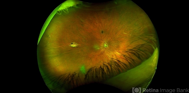
- 66-year-old white male with Gardner Syndrome (colon resection in 1991), who has two children with Gardner Syndrome, presented to Dr. Zocchi with an RD in the fellow eye that was successfully repaired with a pneumatic retinopexy. Currently 20/20 OU with IOP of 7 OD and 14 OS; no RAPD; PCIOL OU. Dr. Zocchi got oral permission by the patient to have these put into the Retina Image Bank. Although the CHRPE like lesions (2 OD) are not bilateral, we both think these lesions represent "retinal pigment epithelial hamartomas associated with familial adenomatous polyposis (RPEH-FAP)" as Shields described in their Intraocular Tumors book. One lesion is located superiorly and is pigmented with depigmented margins; the temporal lesion is atrophic with minimal remaining pigment hypertrophy.


 Associated with Gardner's Syndrome")

")
")


