Search results (135 results)
-
 Meridional Fold
Nov 9 2012 by Norman Byer
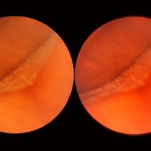
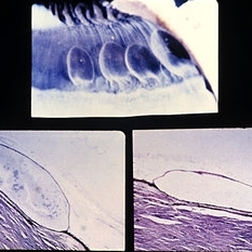
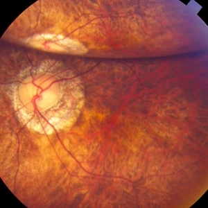
The next two photographs are of the same lesion in a 28-year-old woman. This view shows a sloping retinal mound with a radial retinal fold in the center. This is not a typical meridional fold for it stops short of the ora serrata and there is no dentate process. The upper temporal ora serrata and pars plana are well shown and peripheral cystoid degeneration is present posterior to the ora.
Condition/keywords: ora serrata, pars plana, peripheral cystoid degeneration, radial retinal fold, sloping retinal mound
-
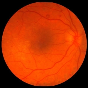
 Pars Plana Cyst Mimicking a Retinal Detachment or Retinoschisis
Apr 8 2021 by Vishak J. John, MD
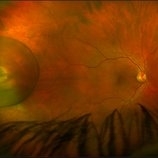
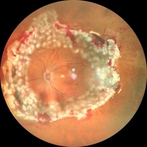
Fundus photo of a 49-year-old male with a large pars plana cyst around an area of chorioretinal scarring.
Photographer: Danielle Lombardo, Vistar Retina Consultants, Roanoke, VA
Imaging device: Optos
Condition/keywords: cyst, pars plana
-
 Pars Plana Snowbank
Oct 9 2012 by Jeffrey G. Gross, MD, FASRS
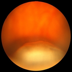
Pars plana snowbank.
Condition/keywords: pars plana, snowbank
-
 Slide 2-36
Feb 19 2019 by Lancaster Course in Ophthalmology
Involvement of the pars plana region in a 55-year-old woman with sarcoid. "Snow bank" exudate over the ciliary body and large vitreous opacities resemble those in the clinical entity "pars planitis."
Condition/keywords: ciliary, pars plana, pars planitis, sarcoid, vitreous opacity
-
 Slide 8-15
Mar 4 2019 by Lancaster Course in Ophthalmology
Retinal detachment due to vitreous incarceration in the cataract wound and traction on the retina inferiorly. The tented-up retina has been pulled over the pars plana. (E.P. No. 32421)
Condition/keywords: cataract, inferiorly, pars plana, vitreous
-
 Slide 9-57
Feb 26 2019 by Lancaster Course in Ophthalmology
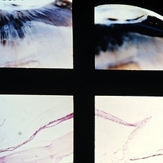
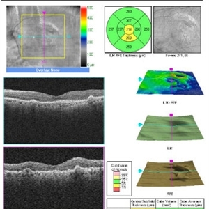
Pars plana cysts. Colloidal iron staining without (lower left) and with (lower right) hyaluronidase demonstrates the presence of hyaluronic acid.
Condition/keywords: hyaluronic acid, pars plana
-
 Slide 9-70
Feb 26 2019 by Lancaster Course in Ophthalmology
Peripheral retinal zonular traction tuft. A strand of fibroglial tissue extends anteriorly over the pars plana from the peripheral retina. A zonular fiber or condensed vitreous strand (arrow) is attached to the apex of the tuft.
Condition/keywords: fibroglial tissue, pars plana, retinal zonular traction tuft
-
 Encircling Buckle Effect
Jul 7 2015 by Hamid Ahmadieh, MD
Late FA image of the right eye of a 30-year-old man who underwent pars plana vitrectomy , endolaser photocoagulation and an encircling band placement a couple of years before following a penetrating trauma at the vitreous base area at the 7 o'clock meridian.
Photographer: Nayereh Hadipour, Negah Eye Center,Tehran, Iran
Imaging device: Specteralis
Condition/keywords: pars plana vitrectomy (PPV)
-
 Fibrosis and Traction Following Traction Retinal Detachment Repair
Oct 13 2020 by Sophia El Hamichi, MD
A 29-year-old female with a history of diabetes mellitus type 1, presented with proliferative diabetic retinopathy OU and tractional retinal detachment OD. The patient underwent retinal detachment repair with pars plana vitrectomy, endolaser and silicone oil placement. After one month of her surgery, the patient presented with retinal fibrosis and tractions depicted in the image.
Photographer: Belinda Rodriguez, Murray Ocular Oncology and Retina, Miami
Condition/keywords: pars plana vitrectomy (PPV), post-op, proliferative diabetic retinopathy (PDR), proliferative vitreoretinopathy (PVR), tractional retinal detachment
-
 Pars Plana Cysts
Jul 29 2020 by Vinod Kumar
Pars plana cysts found during peripheral indentation at the conclusion of vitrectomy for rhegmatogenous retinal detachment.
Photographer: Vinod Kumar
Imaging device: Still from a video
Condition/keywords: cyst of the pars plana
-
 Pars Plana Cysts
Jan 29 2018 by Shani Pillar
During a pars plana vitrectomy for fixation of a dislocated IOL, this finding of pars plana cysts was seen, while performing indentation. Pars plana cysts are not uncommon, but rarely visualized so clearly, given their extremely peripheral location.
Photographer: Dr. Shani Pillar, Meir Medical Center, Kfar Saba, Israel
Imaging device: Intraoperative microscope
Condition/keywords: cyst of the pars plana, ora serrata, peripheral fundus lesion
-
 Pars Plana Vitrectomy TPA 2
Oct 29 2012 by John S. King, MD
About 2 months after PPV, sub-retinal tPA, for the second time a large sub-retinal hemorrhage appeared. Visual acuity 20/40-20/50 after second ppv c subretinal tpa (vs cf prior to PPV), but less than result following first surgery (20/30).
Photographer: Kristin Konecki, OcuSight Eye Care Center, Rochester, NY
Imaging device: Cirrus
Condition/keywords: subretinal hemorrhage
-
 Persistent Fetal Vasculature
Sep 23 2024 by Carlos Augusto Moreira, MD, PhD
Persistent Fetal Vasculature - an intraocular cotton swab appearance.
Photographer: Carlos Augusto Moreira-Neto, Hospital de Olhos do Paraná
Imaging device: NGENUITY Visualization System
Condition/keywords: pars plana vitrectomy (PPV), persistent fetal vasculature (PFV)
-
 Proliferative Diabetic Retinopathy with Severe Subhyaloid Hemorrhage
Oct 15 2012 by Jeffrey G. Gross, MD, FASRS
PDR with severe subhyaloid hemorrhage post-op, PPV.
Condition/keywords: pars plana vitrectomy (PPV), post-op, subhyaloid hemorrhage
-
 Proliferative Vitreoretinopathy
Jun 11 2023 by Ethan K Sobol, MD
Intraoperative view of inferior PVR prior to successful retinal re-attachment
Condition/keywords: pars plana vitrectomy (PPV), proliferative vitreoretinopathy (PVR)
-
 Proliferative Vitreoretinopathy
Jun 11 2023 by Ethan K Sobol, MD
Intraoperative view of a retinal detachment with extensive inferior proliferative vitreoretinopathy, prior to successful retinal re-attachment.
Condition/keywords: pars plana vitrectomy (PPV), proliferative vitreoretinopathy (PVR)
-
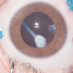
 Reverse Reverse Hypopyon
Jul 15 2019 by Anfisa Ayalon, MD
Slit-lamp photograph of a pseudo-hypopyon of perfluorocarbon in the eye of a young patient who had undergone repair of rhegmatogenous retinal detachment years prior. Note the emulsified appearance.
Photographer: Anfisa Ayalon, MD. Meir Medical Center, Kfar Saba, Israel.
Condition/keywords: pars plana vitrectomy (PPV), perfluorocarbon fluid, retained perfluorocarbon
-
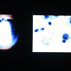 Slide 8-27
Mar 4 2019 by Lancaster Course in Ophthalmology
Ocular reticulum-cell sarcoma. Appearance of vitreous cells and debris (left) in a 61-year-old man with a 2-year history of recalcitrant uveitis. Pars plana vitrectomy specimen was prepared, using a millipore filter and staining by a modified Papanicolaou technique. Tumor cells with nuclear cytoplasmic disproportion and finger like nuclear extensions are characteristic of reticulum-cell sarcoma (right). (E.P. No. 38606)
Condition/keywords: pars plana vitrectomy (PPV), reticulum cell sarcoma, uveitis, vitreous cells
-
 Tools & Techniques for 23, 25, and 27G PPV
Dec 10 2012 by Yale L. Fisher, MD
Dr. Steve Charles reviews the tools and techniques he uses for 23G, 25G, and 27G PPV. NOTE: A narration by Dr. Steve Charles will soon be available for this movie- please check back periodically.
Condition/keywords: pars plana vitrectomy (PPV), video
-
 Vitreous Incarceration
Dec 10 2012 by Yale L. Fisher, MD
Pars plana incision site endoscopic imaging of vitreous incarceration from Dr. Yale Fisher's collection.
Condition/keywords: video
-
 "Internal Mirroring" Effect by Intraocular Gas
Mar 25 2014 by Homayoun Tabandeh, MD, FASRS
"Internal mirroring" by residual intraocular gas in a highly myopic patient 3 weeks post repair of retinal detachment with pars plana vitrectomy and C3F8 gas.
Photographer: Danny Rivas
Condition/keywords: high myopia, intraocular gas
-
25 Gauge Vitrectomy Membrane Shaving
Jan 31 2015 by Thomas A. Ciulla, MD, MBA, FASRS
Membrane shaving of dense membranes in diabetic traction detachment using 25 gauge vitrectomy.
Condition/keywords: diabetes, pars plana vitrectomy (PPV), retina surgery, tractional retinal detachment, vitreoretinal surgery
-
 360 Degree Retinectomy
Sep 11 2020 by Sham Talati, DOMS
A case of retinal detachment with PVR. Patient underwent pars plana vitrectomy with silicon oil injection with 360 degree retinectomy.
Photographer: Dr. Sham Talati,Retina Foundation,Ahmedabad
Imaging device: Nidek Mirante
Condition/keywords: proliferative vitreoretinopathy (PVR), retinectomy
-
 4 Point Scleral Fixation Akreos AO60 With Gore Tex Suture
May 20 2021 by Jesus Lozano, MD
Optos Silverstone fundus image of a 54-year-old man after 4 point scleral fixation Akreos AO60 with Gore Tex suture plus PPV who had a severe traumatic iris defect and was aphakic after ocular trauma.
Photographer: Yair Bet Yosef, Hadassah Medical Center. Israel
Imaging device: Optos Silverstone
Condition/keywords: aphakia, globe perforation, lens, pars plana vitrectomy (PPV), penetrating trauma, vitreous hemorrhage
-
 Aniridic Fibrosis Syndrome - #1 of 7
Jan 24 2013 by Christopher D. Riemann, MD
6-year-old pseudophakic girl with aniridic fibrosis syndrome. Superior view with HD endoscope. Note: complete absence of fibrosis, a normal ciliary body, normal pars plana and normal anterior retina.
Photographer: Christopher Riemann MD, Cincinnati Eye Institute, University of Cincinnati
Imaging device: Endoscope
Condition/keywords: aniridia, epiciliary membrane
 Loading…