Search results (3700 results)
-
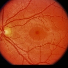
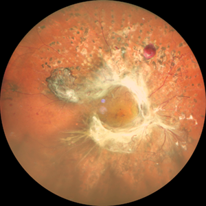
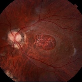 Giant Persistent Macular Hole
Dec 6 2024 by César Adrián Gómez Valdivia, MD
Giant Persistent Macular Hole found in a 48 year-old male patient one year after vitrectomy.
Photographer: @eyemissu2
Imaging device: TOPCON TRC-50DX
Condition/keywords: macular, macular hole
-
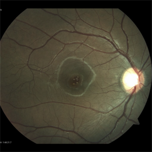
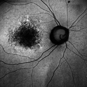
 Macular Hole
Dec 19 2021 by Eduardo Javier Pinuer Alvarado
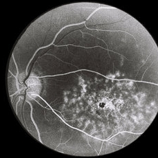
Fundus photograph of a 62-year-old woman with macular hole grade 3-4.
Photographer: Eduardo Pinuer A, Universidad Austral de Chile.
Imaging device: CR-2 AF Digital Non-Mydriatic Retinal Camera, Canon.
Condition/keywords: atrophic retinal hole, macular, retina
-
 Morning Glory Syndrome
Jan 6 2020 by Olivia Rainey
Ultra-wide field pseudocolor image of a 23-month-old male with morning glory syndrome affecting his left eye. Patient presented with esotropia affecting his left eye and strabismic amblyopia affecting both eyes. He could fix and follow on exam and his medical history was unremarkable.
Photographer: Olivia Rainey
Imaging device: Optos California
Condition/keywords: esotropia, left eye, macular, Morning Glory Syndrome, Optos, strabismic amblyopia, ultra-wide field imaging
-
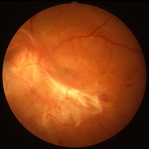 PDR with Traction RD of Macular
Oct 8 2012 by Jeffrey G. Gross, MD, FASRS
PRD, with traction RD of macular, pre-op, 20/200.
Condition/keywords: 20/200, macular, pre-op, tractional retinal detachment
-
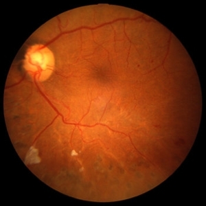 PDR with Traction RD of Macular
Oct 8 2012 by Jeffrey G. Gross, MD, FASRS
PDR with traction RD of macular, post-op, 20/40.
Condition/keywords: 20/40, macular, post-op, tractional retinal detachment
-
 Retinal Microaneurysms & Dot/Blot Hemes Fundus Photo OS
May 12 2025 by Briana Hernandez
OS Optos Fundus Photo of Retinal Microaneurysms & Dot/Blot Hemes in 91-year-old female BRVO patient.
Photographer: Briana Hernandez, Hilton Head Retina Institute
Imaging device: Optos
Condition/keywords: macular
-
 Slide 13-5
Mar 4 2019 by Lancaster Course in Ophthalmology
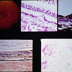
Low-power view of an undifferentiated retinoblastoma underlying the macular area ( x25).
Condition/keywords: macular, retinoblastoma
-
 Slide 2-29
Feb 19 2019 by Lancaster Course in Ophthalmology
Macular area in subacute sclerosing panencephalitis. Retina has wrinkled internal limiting membrane. Most of the retinal layers are destroyed. Two large, red intranuclear inclusions are visible, in the mid-retinal cells, on the left and right. Note the abrupt loss of RPE under the area and the minimal number of lymphocytes.
Condition/keywords: intranuclear, lymphocytes, macular, retinal pigment epithelium, subacute sclerosing panencephalitis
-
 Slide 9-85
Feb 26 2019 by Lancaster Course in Ophthalmology
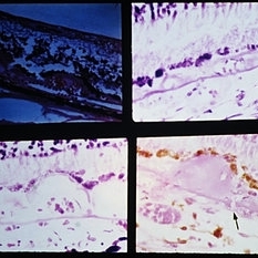
Drusen in the macular area. They are periodic-acid-Schiff-positive (upper and lower right), and stain positive for lipid with the oil red-O technique (lower left). The overlying RPE is effaced, as shown in routine section (upper right) and by flat preparation (lower right).
Condition/keywords: drusen, macular, retinal pigment epithelium
-
 Slide 9-88
Feb 26 2019 by Lancaster Course in Ophthalmology
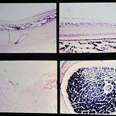
Vascularized drusen in periphery (upper left) and macular area (upper right and lower left). Note the tiny break in Bruch's membrane (arrow) with a choroidal vessel with an erythrocyte extending into a peripapillary druse.
Condition/keywords: Bruch's membrane, drusen, macular
-
 Slide 9-89
Feb 26 2019 by Lancaster Course in Ophthalmology
Nutritional amblyopia. The nerve fiber and ganglion cell layers are absent in the macular area (upper views). The temporal side of the optic nerve head (lower left) is partially atrophy, with marked reduction in the size of the nerve fiber bundles and secondary gliosis.
Condition/keywords: amblyopia, atrophy, gliosis, macular
-
 Solar Retinopathy
Feb 12 2015 by H. Michael Lambert, MD
Yellow white spot on central fovea, serous detachment on macular.
Condition/keywords: macular, solar retinopathy
-
 Solar Retinopathy
Feb 12 2015 by H. Michael Lambert, MD
Yellow white spot on central fovea, serous detachment on macular.
Condition/keywords: macular, solar retinopathy
-
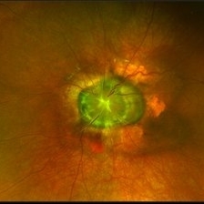
 Thickness Macular Hole
Apr 10 2024 by Tareq Alsulami
Fundus photography of an 14 years old man with Full Thickness Macular Hole after direct exposure of the blue laser to his eye
Photographer: Tareq alsulami ophthalmic technician-king Abdullah Medical City-Saudi Arabia
Imaging device: ZEISS
Condition/keywords: holes, macular
-
 27-Gauge Vitrectomy
Aug 3 2013 by Yusuke Oshima, MD, PhD
A high-performance 27-gauge vitrectomy system (27+) was used for treating a case with primary rhegmatogenous retinal detachment complicated with macular hole.
Condition/keywords: macular hole, video, vitrectomy
-
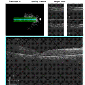
 5 Weeks Post JETREA
May 15 2013 by Robert T. Wendel, MD
5 weeks post-op JETREA for stage 2 MH. OCT shows closing macular hole, but the cortex still attached.
Condition/keywords: macular hole, optical coherence tomography (OCT)
-
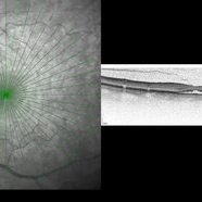
 72 hours macular OCT post blunt trauma. Impending macular hole.
Dec 29 2012 by Humberto Ruiz-Garcia, MD
Macular OCT image showing impending macular hole posterior to blunt trauma.
Photographer: Humberto Ruiz-Garcia
Imaging device: Cirrus HD OCT
Condition/keywords: macular hole
-
---thumb.jpg/image-square;max$300,300.ImageHandler) Acute retinal necrosis
Acute retinal necrosis
Feb 15 2013 by From the Collections of Thomas M. Aaberg, MD and Thomas M. Aaberg Jr., MD
Diffuse intraretinal hemorrhages and whitening in the posterior pole consistent with acute retinal necrosis.
Condition/keywords: macular edema, microangiopathy, retinal necrosis, retinal whitening
-
 Aggressive Posterior Retinopathy of Prematurity with Macular Hemorrhage
Oct 9 2012 by Audina M. Berrocal, MD FASRS
APROP with multiple pre-retinal hemorrhages
Photographer: Ditte Hess CRA, BPEI
Imaging device: RETCAM
Condition/keywords: macular hemorrhage, retinopathy of prematurity (ROP)
-
 Annular Tractional Retinal Detachment
Jul 4 2024 by Hector Gabriel Moreno Solano, MD, MHA
52-year-old Hispanic female patient with a diagnosis of type II diabetes mellitus of 15 years of evolution, comes to the retina service for progressive visual loss in the right eye (single functional eye) with visual acuity of 20/100, Fundus examination reveals laser-modified proliferative diabetic retinopathy with activity + annular tractional retinal detachment with macular involvement.
Photographer: Hector Gabriel Moreno Solano, MD, MHA, HGZ #20 IMSS Puebla.
Imaging device: Mirante
Condition/keywords: macular detachment, proliferative diabetic retinopathy (PDR), tractional retinal detachment
-
 Autoflluorescence in Macular Dystrophy
Jun 10 2020 by Manish Nagpal, MD, FRCS (UK), FASRS
Autofluorescence in a case of macular dystrophy.
Photographer: Gayathri Mohan, Retina Foundation
Imaging device: NIDEK SLO MIRANTE
Condition/keywords: macular dystrophy
-
---thumb.JPG/image-square;max$300,300.ImageHandler) Behcet's Disease
Behcet's Disease
Nov 25 2012 by Mallika Goyal, MD
Fundus photograph of right eye of a 23-year-old gentleman with Behcet's Disease shows occlusive retinal vasculitis with optic disc pallor and macular ischaemia. Other eye has similar appearance with no light perception.
Photographer: Mallika Goyal, MD, Apollo Health City, Hyderabad, India
Condition/keywords: macular ischemia, occlusive vasculitis
-
 Behcet's Disease
Nov 25 2012 by Mallika Goyal, MD
Fundus photograph of left eye of a 23-year-old gentleman with Behcet's Disease shows occlusive retinal vasculitis with optic disc pallor and macular ischemia. This eye has no light perception; other eye has similar fundus appearance.
Photographer: Mallika Goyal, MD, Apollo Health City, Hyderabad, India
Condition/keywords: macular ischemia, occlusive vasculitis, optic disc pallor
-
 Berlin's Edema
May 2 2013 by Henry J. Kaplan, MD
Macular Berlin's edema following blunt trauma.
Condition/keywords: Berlin's edema
-
 Best Disease
Apr 24 2024 by Marcelo Zas, MD PhD
Best vitelliform macular dystrophy (BVMD) or Best disease. Is the most common autosomal dominant macular dystrophy. It involves the retinal pigment epithelium (RPE), and leads to a characteristic bilateral yellow “egg-yolk” appearance of the macula as you can see in this image. Essentially, BVMD is considered to have 6 clinical stages: Previtelliform, Vitelliform, Pseudohypopyon, Vitelleruptive, Atrophic and Choroidal neovascularization. As the disease progresses, patients may experience a slow, bilateral decrease in visual acuity, central scotoma, or metamorphopsia. With secondary CNV, visual decline can be rapid, however.
Photographer: Luciano Scorsetti MD
Condition/keywords: Macular Dystrophy
 Loading…