Search results (3700 results)
-
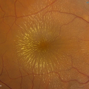 Cat Scratch
Feb 15 2017 by Hua Gao, MD, PhD, FASRS
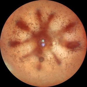
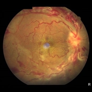
A female patient of 57-year-old presented with neuroretinitis due to cat-scratch disease with positive Bartonella henselae antibodies. Two weeks after symptom onset she developed exudates in a "macular star" pattern.
Condition/keywords: cat scratch retinitis
-
 Retinal Detachment with PVR (s/ SPR, PPV, MPV, 360 Retinectomy, PFO, PI, FAx, SO)
Aug 22 2019 by Merrick Avila
Ultra-wide field pseudocolor fundus photograph of a 64-year-old female with a treated retinal detachment with proliferative vitreoretinopathy. Patient has a history of complex retinal detachments that have been treated multiple times. On exam 8-22-19, there were large macular holes with LP vision. There was a long discussion about guarded nature of her condition and goals or trial for repair including globe sparing prevention of phthisis.
Photographer: Merrick Avila
Imaging device: Optos
Condition/keywords: diabetic retinopathy, hemorrhage, Optos, proliferative vitreoretinopathy (PVR), retinectomy, silicone oil
-
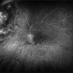
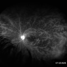 Retinal Vasculitis in Behcet's OS
Jun 29 2018 by Gareth Lema, MD, PhD
IVFA at 7 minutes showing retinal vasculitis, cystoid macular edema, and disc staining.
Photographer: Ross Eye Institute, University at Buffalo Jacobs School of Medicine, Buffalo. NY
Imaging device: Optos
Condition/keywords: Behcet's Disease, cystoid macular edema (CME), disc staining, retinal vasculitis
-
 Acute Macular Neuroretinopathy
Dec 11 2019 by Lauren Whaley
34-year-old female patient presented with changes in vision after recent upper respiratory infection. Referring doctor originally thought it was a blood pressure issue. She noticed a "C" shape in her vision. Infrared image was captured showing exactly what patient was describing! Doctor confirmed with this image that it was AMN.
Photographer: Lauren R. Whaley, COA
Imaging device: Heidelberg Spectralis
Condition/keywords: 30 degrees, acute macular neuroretinopathy, Heidelburg Spectralis, left eye, macula, near infrared autofluorescence (NIRAF)
-
 Choroidal Fracture
Oct 27 2024 by César Adrián Gómez Valdivia, MD
Fundus photograph of a traumatic choroidal fracture & extra-macular sub-retinal hemorrhage.
Photographer: @eyemissu2
Imaging device: TOPCON TRC-50DX
Condition/keywords: Choroidal Fracture
-
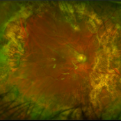
 Cuticular and soft drusen
Jun 14 2021 by Gerardo Garcia-Aguirre, MD
Fundus photograph (left) and Retro mode infrared image (right) of an eye with soft and cuticular drusen. Drusen are highlighted and better visualized with retro mode imaging.
Photographer: Gerardo Garcia-Aguirre
Imaging device: Nidek Mirante
Condition/keywords: drusen, dry age-related macular degeneration (dry AMD)
-
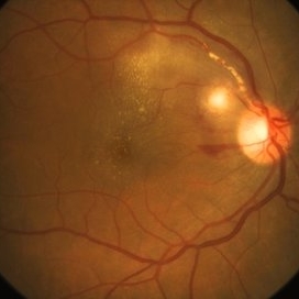
 Epiretinal Membrane/Macular Pucker With Combined Hamartoma of Retina and RPE
Jul 8 2015 by Emmanuel Chang, MD PhD FACS FASRS
10-year-old with history of progressive severe distortion in the left eye over the past year.
Photographer: Retina and Vitreous of Texas
Imaging device: Heidelberg Autofluorescence
Condition/keywords: combined hamartoma, epiretinal membrane (ERM), retinal pigment epithelium (RPE) hamartoma
-
 Green Goblin Detachment
Jan 13 2022 by Netan Choudhry, MD, FRCS(C) FASRS
Tractional retinal detachment with macular hole in a 76-year-old female.
Photographer: John Golding BA, Vitreous Retina Macula Specialists of Toronto, OCTane Imaging Lab
Imaging device: Multicolor fundus photo taken on the Spectralis OCT2 (Heidelberg Engineering GmbH).
Condition/keywords: macular hole, Multispectral imaging, tractional retinal detachment
-
 Hypertensive Retinopathy
Feb 25 2013 by Suber S. Huang, MD, MBA, FASRS
32-year-old African American male with Grade IV hypertensive retinopathy and acute renal failure. Vision OD 20/70, OS 20/25. Creatine 7.1. BP: 250/150.
Photographer: Geoffrey Pankhurst, University Hospitals, Eye Institute/Dept. Ophthalmology and Visual Sciences Case Western Reserve University Cleveland, OH
Imaging device: Topcon TRC 50x
Condition/keywords: acute renal failure, disc edema, exudate, hypertension, hypertensive retinopathy, ischemia, macular edema, macular ischemia, optic disc edema
-
 Lady in a dress
Feb 9 2023 by Shelby Helton
Fluorescein Angiography on a 67-year-old male with significant RPE changes secondary to a severe subretinal hemorrhage that required a vitrectomy with subretinal TPA in 2013.
Photographer: Shelby Helton
Imaging device: Heidelberg Spectralis
Condition/keywords: wet age-related macular degeneration (wet AMD)
-
 Myopic Traction Maculopathy
Mar 17 2025 by Drew Mitchell
HD 1 line 100x 9 mm scan of a right eye with MTM at stage 3c. Macular Schisis Detachment.
Photographer: Drew Mitchell OCT-C
Imaging device: Zeiss Cirrus 5000
Condition/keywords: full thickness macular hole, Macular hole, myopic foveoschisis, myopic macular schisis, myopic traction maculopathy, PVD
-
 Proliferative Diabetic Retinopathy
Jul 15 2022 by Gabriel Costa Andrade, PhD
Fundus angiography of an 22-year-old man with proliferative diabetic retinopathy and macular ischemia.
Photographer: Dr Gabriel Andrade
Condition/keywords: Diabetes
-
 Radiation Retinopathy; BRVO with Macular Edema
Apr 26 2023 by Denica Rodriguez
Ultra-wide field fluorescein angiography of a 61 year old male with radiation retinopathy following brachytherapy for choroidal melanoma of his left eye. Following treatment, patient developed a branch retinal vein occlusion both ischemic and non-ischemic. Anti-VEGF injections were recommended. The fine needle biopsy showed a class 2 uveal melanoma. Patient also has diabetic retinopathy affecting both eyes. Patient's vision at the time the image was taken was Dcc 20/80-1.
Photographer: Denica Rodriguez COA, ST
Imaging device: Optos California
Condition/keywords: branch retinal vein occlusion (BRVO), Choroidal melanoma, diabetic retinopathy, FA, fluorescein angiogram (FA), I-125 brachytherapy, macular edema, melanoma, Optos, radiation retinopathy, Retina, ultra-wide field imaging
-
 Retinitis Pigmentosa with Macular Hole with Posterior Subcapsular Cataract
Apr 28 2025 by Malvika Singh
Fundus photograph of the right eye of a 31 year old with retinitis pigmentosa with a macular hole, showing the shadow of posterior subcapsular cataract over the fundus.
Photographer: Dr Malvika Singh, Retina Foundation, Ahmedabad, India
Imaging device: Mirante SLO/OCT
Condition/keywords: macular hole, posterior subcapsular cataract, retinitis pigmentosa
-
 Submacular PFO
Feb 20 2020 by Kevin J. Blinder, MD, FASRS
This is a 53-year-old gentleman that was referred to us for a second opinion with an inoperable RD with PVR after 3 failed attempts. We performed a PPV, membranectomy, scleral buckling procedure, with silicone oil injection. This case did not require PFO. You can imagine our surprise when we discovered submacular PFO postoperatively. It is very difficult to see the PFO on the Optos. The infrared shows it clearly, with confirmation of the submacular space on the SD-OCT.
Photographer: Jarrod Wehmeier, The Retina Institute; St. Louis, MO
Imaging device: optos
Condition/keywords: submacular perfluorocarbon liquid (PFO)
-
 Aggressive Posterior Retinopathy of Prematurity with Macular Hemorrhage
Oct 9 2012 by Audina M. Berrocal, MD FASRS
APROP with multiple pre-retinal hemorrhages
Photographer: Ditte Hess CRA, BPEI
Imaging device: RETCAM
Condition/keywords: macular hemorrhage, retinopathy of prematurity (ROP)
-
 Benign Idiopathic Hemorrhagic Retiopathy - RF Image
Feb 6 2013 by Roy Schwartz, MD
A healthy 31-year-old female presented with visual deterioration in her left eye. On examination thick macular retinal hemorrhages were seen, as well as some small minopr hemorrhages in the periphery. A workup for hypercoagulability was negative, and sources of emboli were not found. A diagnosis of a unilateral idiopathic hemorrhagic retinopathy was made.
Photographer: Galit Yair-Pur
Condition/keywords: benign idiopathic hemorrhagic retinopathy, red-free
-
 Branch Retinal Vein Occlusion with Macular Edema
Aug 23 2012 by Gerardo Garcia-Aguirre, MD
Fundus photograph composition of the left eye, showing flame-shaped and blot hemorrhages in the superotemporal quadrant, with hard exudates surrounding the fovea.
Photographer: Noemí Hernández, Asociación para Evitar la Ceguera en México
Condition/keywords: branch retinal vein occlusion (BRVO), macular edema
-
 Cat Scratch Disease
Mar 29 2021 by Gabriel Costa Andrade, PhD
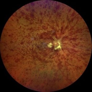
Fundus photograph of an 36-year-old woman with a macular vasculitis, pre retinal hemorrhage and exudation due to Bartonella henselae infection.
Photographer: Gabriel Andrade
Condition/keywords: cat scratch retinitis
-
 Central Retinal Vein Occlusion
Jun 21 2025 by Moazzam Parvez
Fundus photograph of a 56 year old male presenting with dilated tortuous vessels with adjoining Hard exudates and macular star.
Photographer: Moazzam Parvez , Netralayam , Kolkata
Imaging device: Topcon Maestro 2
Condition/keywords: CRVO with macular edema, hard exudates, macular star
-
 Central Retinal Vein Occlusion associated with disc edema
Oct 19 2023 by Gabriel Costa Andrade, PhD
53-year-old woman with an acute CRVO. The patient has a history of breast cancer undergoing treatment with systemic chemotherapy. Notice the peripapillary cotton wool spots, superficial flame shaped hemorrhages and deeper dot and blot hemorrhages in all 4 quadrants.
Photographer: Gabriel Andrade
Condition/keywords: central retinal vein occlusion (CRVO), macular edema, Retina
-
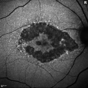
 Cuticular Drusen
Jan 17 2024 by John Lee
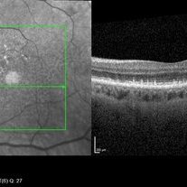
Heidelberg SD-OCT of a 65-year-old woman with age-related macular degeneration demonstrating classic sawtooth appearance of cuticular drusen.
Photographer: Natasha Vinson
Imaging device: Heidelberg Spectralis
Condition/keywords: age-related macular degeneration (AMD), cuticular drusen
-
 Dry AMD
Jun 4 2014 by Henry J. Kaplan, MD
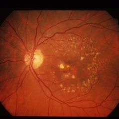
Multiple drusen with RPE changes in the macula #2.
Condition/keywords: age-related macular degeneration (AMD), dry age-related macular degeneration (dry AMD)
-
 Dry AMD, Advanced Atrophic without Subfoveal Involvement
Oct 12 2021 by Kelli Nyenhuis
OCT Heidelberg photograph of a 79-year-old woman with AMD advancing that has been observed over the last 10 years.
Photographer: Kelli Nyenhuis, OMA
Imaging device: Heidelberg
Condition/keywords: dry age-related macular degeneration (dry AMD)
-
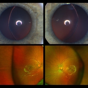 Ectopia Lentis
Jan 21 2021 by Jamin S. Brown, MD
This image serial demonstrates a patient with simple ectopia lentis. Anterior segment photographs in the upper panel demonstrate nasally subluxated crystalline lenses. Widefield fundus photography shows a "pseudo-buckle" which is the result of an optical effect due to the lens subluxation (artifactual image enlargement). Also note the juvenile macular reflex in this young patient. Ectopia lentis can present isolated ("simple") or in combination with various systemic defects (Marfan's syndrome, Weil-Marchesani syndrome or Ehlers-Danlos syndrome to name a few). Isolated ectopia lentis can be hereditary and causative genes have been identified as ADAMTSL4 located on chromosome 4 and FBN1 gene located on chromosome 15. Defects in the genes cause weakness in the zonular fibers which can lead to lens dislocation. Lastly, various ocular disorders such as Aniridia, Axenfeld-Rieger, Pseudoexfoliation or Trauma may also result in lens dislocation or subluxation.
Photographer: Stefanie Palmer CRA, Retina Vitreous Surgeons of CNY
Condition/keywords: dislocated lens, ectopia lentis
 Loading…