Search results (3700 results)
-
 Anastomosis
Jul 29 2025 by Drew Mitchell
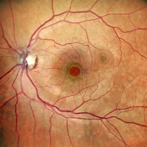
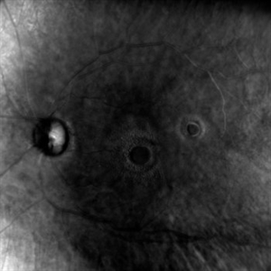
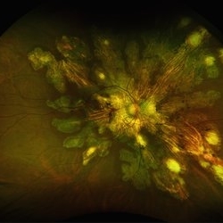
3x3 OCT-Angiography Full Depth Color Coded of a left eye with Macular Telangiectasia Type 2
Photographer: Drew Mitchell, OCT-C
Imaging device: Zeiss Cirrus 5000
Condition/keywords: chorioretinal anastomosis, macular telangiectasia type 2, retinochoroidal anastomosis
-
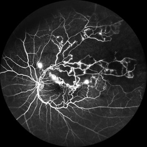 Branch Retinal Vein Occlusion
Jul 23 2025 by Malvika Singh
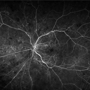
Fluorescein angiogram of a 52 year old man showing capillary non perfusion areas and leakages along the superotemporal arcade and at the macula.
Photographer: Dr Malvika Singh, Retina Foundation, Ahmedabad, India
Imaging device: Mirante SLO/OCT
Condition/keywords: branch retinal vein occlusion (BRVO), CNP areas, FLUORESCEIN ANGIOGRAPHY, fluorescein leakage, macular edema
-
 Neuroretinitis
Jul 7 2025 by César Adrián Gómez Valdivia, MD
Neuroretinitis found in a 38 YO male patient with IV drugs abuse history. Findings were bilateral. The lipid-rich component of the exudate is able to penetrate into the outer plexiform layer, creating what is clinically seen as a macular star pattern.
Photographer: @eyemissu2
Imaging device: TOPCON TRC-50DX
Condition/keywords: neuroretinitis
-
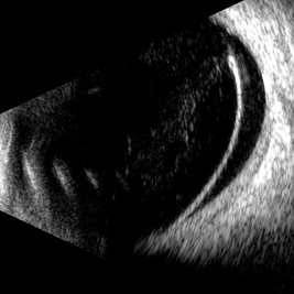 Retinal Dialysis
Jul 5 2025 by Gustavo Uriel Fonseca Aguirre
This B-mode longitudinal ultrasound scan demonstrates a retinal dialysis, appearing as a linear discontinuity at the ora serrata with associated vitreous base avulsion. The scan reveals mild subretinal fluid extending from the dialysis site with macular involvement.
Photographer: Gustavo U. Fonseca Aguirre, Hospital Conde de Valenciana, Ciudad de México
Condition/keywords: retinal dialysis
-
 Giant Persistent Macular Hole
Jul 5 2025 by César Adrián Gómez Valdivia, MD
Giant Persistent Macular Hole found in a 48YO male patient 1 year after vitrectomy.
Photographer: @eyemissu2
Imaging device: TOPCON TRC-50DX
Condition/keywords: macular hole
-
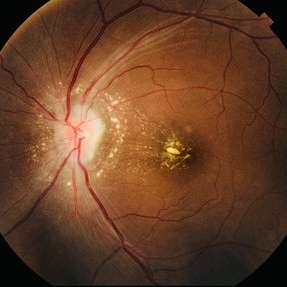
 Retinal Vein Occlusion
Jul 5 2025 by T. P . VIGNESH, MBBS,MS
Fundus photograph of a 52-year-old man with a macular branch retinal vein occlusion and macular edema.
Photographer: Sivanath
Imaging device: EIDON
Condition/keywords: branch retinal vein occlusion (BRVO)
-
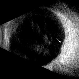 Diabetic Macular Edema
Jul 3 2025 by Gustavo Uriel Fonseca Aguirre
This B-mode longitudinal ultrasound scan demonstrates diabetic macular edema with mild subretinal fluid accumulation, appearing as a subtle hypoechoic space beneath the neurosensory retina. The macular region shows retinal thickening and heterogeneous medium reflectivity, consistent with active exudative changes (arrow). No vitreomacular traction is observed.
Photographer: Gustavo U. Fonseca Aguirre, Hospital Conde de Valenciana, Ciudad de México
Condition/keywords: diabetic macular edema
-
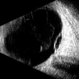 Macular Retinoschisis
Jul 3 2025 by Gustavo Uriel Fonseca Aguirre
This B-mode longitudinal ultrasound scan reveals macular retinoschisis, demonstrating a characteristic splitting of retinal layers with a smooth, dome-shaped elevation. The lesion shows maintained structural integrity of both inner and outer retinal walls without associated subretinal fluid or vitreous traction.
Photographer: Gustavo U. Fonseca Aguirre, Hospital Conde de Valenciana, Ciudad de México
Condition/keywords: macular retinoschisis
-
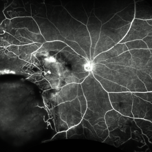 Fluorescein Angiography (FA) of a Primary Retinal Vasoproliferative Tumor
Jun 29 2025 by Marcelo Zas, MD PhD
We present a case of a 33-year-old male patient, who presented with decreased visual acuity in his right eye with 20/80, presenting a primary retinal vasoproliferative tumor in the lower temporal quadrant. The fluorescein angiography findings are: 1. Early hyperfluorescence due to its rich intrinsic vascularity and often has dilated feeding arterioles and draining venules. 2. Marked progressive leakage from the tumor vessels. 3. The late leakage often obscures fine vascular details in the late phase and corresponds to exudation and macular edema seen clinically. 4. Staining of surrounding exudates, RPE disturbances and gliosis. 5. In our case also a marked peripheral capillary closure in the areas adjacent to the tumor and in other quadrants as well.
Photographer: Marcelo Zas MD PhD
Condition/keywords: RETINAL VASOPROLIFERATIVE TUMOR
-
 Double Trouble
Jun 28 2025 by Tejaswita Verma
Fundus image of a 60 year old diabetic female with double macular holes with 6/60 vision status post LE PPV+gas 4 months ago. Other eye also had an unoperated large macular hole. Known case of glaucoma
Photographer: Dr. Tejaswita Verma
Imaging device: MIRANTE
Condition/keywords: macular hole
-
 Double Trouble
Jun 28 2025 by Tejaswita Verma
Retro image of aa double macular hole in a 60 yr old diabetic female status post PPV + gas 4 months ago. Vision was 6/60 in LE.
Photographer: Dr. Tejaswita Verma
Imaging device: MIRANTE
Condition/keywords: macular hole, retro mode
-
 BRVO-MCR-FFA
Jun 27 2025 by Gayathri M S
Case of impending Macular BRVO. 52 year old female on medication for Hypertension and Diabetes Mellitus since 2 years. BCVA 6/18,N6. IOP 16 mmHg. Multicolor Reflectance and Fundus Fluorescein Angiography picture shows mild dilated tortuous inferior vessels, small areas of capillary non perfusion and few microanurysms.
Photographer: Gayathri MS
Imaging device: Heidelberg spectralis
Condition/keywords: fluorescein angiogram (FA), macular branch retinal vein occlusion (BRVO), multicolor
-
 Macular Retinoschisis
Jun 26 2025 by rohan jain
Macular retinoschisis
Photographer: Dr. ROHAN JAIN
Condition/keywords: juvenile retinoschisis, RETINOSCHISIS
-
 Macular Retinoschisis
Jun 26 2025 by rohan jain
Macular retinoschisis
Photographer: Dr. ROHAN JAIN
Condition/keywords: inferotemporal retinoschisis, juvenile retinoschisis, Macular retinoschisis, RETINOSCHISIS
-
 Double Macular Holes
Jun 26 2025 by Moazzam Parvez
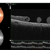
OCT image of a 62 year old man after a blunt trauma by a tennis ball with a vision of CF 3 mt in the right eye.
Photographer: Moazzam Parvez , Netralayam , Kolkata
Imaging device: Topcon Maestro 2
Condition/keywords: double, traumatic macular hole
-
 Two Suns in the Macular Sky
Jun 26 2025 by Moazzam Parvez
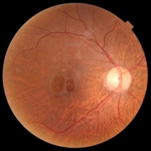
Fundus photograph of a 62 year old gentleman presenting with double adjacent full thickness macular holes in the right eye maintaining a vision of CF 3 mts.
Photographer: Moazzam Parvez ,Netralayam , Kolkata
Imaging device: Topcon Maestro 2
Condition/keywords: double, Macular hole, traumatic macular hole
-
 Retinal Vasoproliferative Tumor
Jun 24 2025 by Marcelo Zas, MD PhD
We present a case of a 33-year-old male patient, who presented with decreased visual acuity in his right eye with 20/80, presenting a primary retinal vasoproliferative tumor in the lower temporal quadrant. The tumor is associated with serous retinal detachment, hard exudation, neovascularization and telangiectasias. Lipid exudates extend toward the macula, indicating macular involvement, which may contribute to decreased visual acuity. Oi was normal with 20/20 of BCVA. The patient was treated initially with IV anti-VEGF therapy and cryotherapy.
Photographer: Marcelo Zas MD PhD
Condition/keywords: RETINAL VASOPROLIFERATIVE TUMOR
-
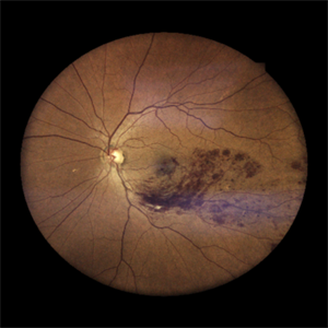
 Serpiginous Choroidopathy
Jun 23 2025 by César Adrián Gómez Valdivia, MD
Fundus photograph of a 29 year-old female patient diagnosed with Serpiginous Choroidopathy. Finings were bilateral. The most common complication of SC is choroidal neovascularization affecting up to 35% of patients. Other reported complications are subretinal fibrosis, cystoid macular edema, branch vein occlusion, serous retinal detachment, optic disc neovascularization ,and anterior uveitis.
Photographer: @eyemissu2
Imaging device: TOPCON TRC-50DX
Condition/keywords: serpiginous choroiditis
-
 Serpiginous Choroidopathy
Jun 23 2025 by César Adrián Gómez Valdivia, MD
Fundus photograph of a 29 year-old female patient diagnosed with Serpiginous Choroidopathy. Finings were bilateral. The most common complication of SC is choroidal neovascularization affecting up to 35% of patients. Other reported complications are subretinal fibrosis, cystoid macular edema, branch vein occlusion, serous retinal detachment, optic disc neovascularization, and anterior uveitis.
Photographer: @eyemissu2
Imaging device: California ICG OPTOS
Condition/keywords: serpiginous choroiditis
-
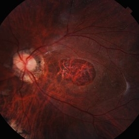
 Central Retinal Vein Occlusion
Jun 21 2025 by Moazzam Parvez
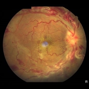
Fundus photograph of a 56 year old male presenting with dilated tortuous vessels with adjoining Hard exudates and macular star.
Photographer: Moazzam Parvez , Netralayam , Kolkata
Imaging device: Topcon Maestro 2
Condition/keywords: CRVO with macular edema, hard exudates, macular star
-
 Central Retinal Vein Occlusion With Waldenstroms macroglobulinemia
Jun 18 2025 by Korey Starkey
64-year-old patient presents with CRVO with secondary macular edema in both eyes. Venous beading present in 2/4 quadrants OU. Patient diagnosed with Waldenstroms macroglobulinemia, found on SPEP and bone marrow biopsy. Treatment recommended of anti-vegF intravitreal injections OU.
Photographer: Korey Starkey
Imaging device: Optos
Condition/keywords: attenuated vessels, central retinal vein occlusion (CRVO), CRVO, FA early phase, FLUORESCEIN ANGIOGRAPHY, macular edema, Optomap, OPTOS CALIFORNIA, severe NPDR, venous beading, Waldenstroms macroglobulinemia
-
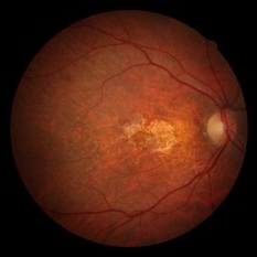
 Extensive Macular Atrophy with Pseudodrusen EMAP OS
Jun 5 2025 by Rogerio N Shinsato, MD, PhD
Extensive Macular Atrophy with Pseudodrusen Patient with EMAP associated with rheumatic fever and benzathine penicillin use. Left eye
Photographer: Rogério N Shinsato
Imaging device: Canon CX-1
Condition/keywords: EMAP
-
 Extensive Macular Atrophy with Pseudodrusen (EMAP) OD
Jun 5 2025 by Rogerio N Shinsato, MD, PhD
Extensive Macular Atrophy with Pseudodrusen Patient with EMAP associated with rheumatic fever and benzathine penicillin use. Right eye
Photographer: Rogério N Shinsato
Imaging device: Canon CX-1
Condition/keywords: macular atrophy
-
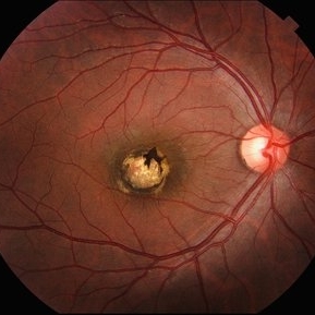 Macular Coloboma
Jun 5 2025 by César Adrián Gómez Valdivia, MD
Macular Coloboma found in a 28 year-old male patient, visual acuity was 20/60. Resulting due to fusion failure of the optic fissure, colobomas are commonly found in the infero-nasal quadrant. If the retina is involved, it is reduced to glial tissue with no underlying RPE or choroid. This appears as an area of whitening often with pigment deposition at the junction of the coloboma and normal retina. Findings were bilateral.
Photographer: @eyemissu2
Imaging device: TOPCON TRC-50DX
Condition/keywords: coloboma
-
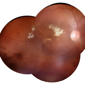 Macular Edema
Jun 4 2025 by Paulina Araujo
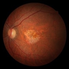
The composite fundus photograph of the right eye demonstrates circinate hard exudates in the thickened macular area, along with flame-shaped intraretinal hemorrhages along the inferior temporal arcade.
Photographer: Paulina D.Araujo Martínez, Asociación para Evitar la Ceguera en México I.A.P., Hospital Dr Luis Sánchez Bulnes.
Condition/keywords: macular edema
 Loading…