Search results (22 results)
-
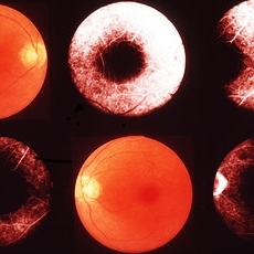
 Cyclitis
Apr 25 2013 by Howard Schatz, MD
23-year-old white female, cyclitis, 20/20; 1/200.
Condition/keywords: cyclitis
-
 Cyclitis
Apr 25 2013 by Howard Schatz, MD
65-year-old white female, III cyclitis, right eye: 20/20; left eye: 20/70.
Condition/keywords: cyclitis
-
 Cyclitis
Apr 25 2013 by Howard Schatz, MD
55-year-old white female, III cyclitis, right eye: 20/64; left eye: 20/320.
Imaging device: Cyclitis
Condition/keywords: cyclitis
-
 Cyclitis
Apr 25 2013 by Howard Schatz, MD
72-year-old white female, III cyclitis, right eye: 20/64; left eye: 20/50.
Condition/keywords: cyclitis
-
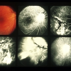
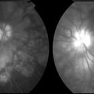
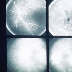 Harada's Disease
May 14 2013 by Howard Schatz, MD
51-year-old white female, 20/100; Harada's disease, cyclitis.
Condition/keywords: cyclitis, Harada's disease
-
 Inflammation
May 16 2013 by Howard Schatz, MD
Prescribed cyclitis, cystoid gone, 20/50; 20/40.
Condition/keywords: cyclitis, cystoid, inflammation
-
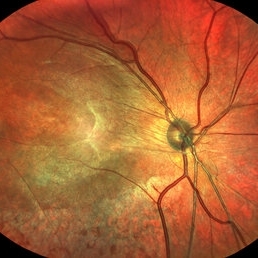 Uveitis
Nov 25 2022 by Filip Kecer
Multicolor composite of an 15 year old boy with cyclitis posterior, first diagnosed with uveitis intermedia retinoschisis o.dx., since then he underwent several procedures, such as PPV, ILM peeling, capsulectomy, retinotomy, transscleral cryo
Photographer: Filip Kecer, National Institute of Childrens Diseases
Imaging device: Spectralis, Heidelberg Engineering
Condition/keywords: cyclitis, epiretinal membrane (ERM), uveitis
-
 ARN (#1) Initial Photo
May 27 2019 by John S. King, MD
60-year-old African American female who had been treated for iridocyclitis for at least a week sent in for vitritis and a nasal fundus lesion. Complaints included redness, floaters, photophobia, and decreased vision. Husband had recent shingles. Acuity was 20/60-2 with IOP of 12, and small KP in Art's triangel, 1-2+ a/c cell, 2-3+ ant vit cell, diffuse arteriolar sheathing, multiple areas of retinal whitening in periphery and mid-periphery (see Photo #1). PCR of a/c was performed, and intravitreal GCV administered, and VACV 2g qid and ASA started.... PCR positive for HZV, pred taper was started two days after presentation as the infection had begun to stablize..... Five days from presentation the vision was 20/60, inflammation and areas of retinal whitening had improved (see Photo #2).... One week later acuity was 20/30, the a/c was quiet and KP resolved; ant vitreous cell decreased; and there was further improvement in retinal appearance without any signs of retinal holes or detachment; she is now on low dose maint VACV (see photo#3)
Photographer: Maysee Yang
Imaging device: Optos CA
Condition/keywords: acute retinal necrosis, Herpes zoster
-
 ARN (#2) Five Days Since Initial Visit
May 27 2019 by John S. King, MD
60-year-old African American female who had been treated for iridocyclitis for at least a week sent in for vitritis and a nasal fundus lesion. Complaints included redness, floaters, photophobia, and decreased vision. Husband had recent shingles. Acuity was 20/60-2 with IOP of 12, and small KP in Art's triangel, 1-2+ a/c cell, 2-3+ ant vit cell, diffuse arteriolar sheathing, multiple areas of retinal whitening in periphery and mid-periphery (see Photo #1). PCR of a/c was performed, and intravitreal GCV administered, and VACV 2g qid and ASA started.... PCR positive for HZV, pred taper was started two days after presentation as the infection had begun to stablize..... Five days from presentation the vision was 20/60, inflammation and areas of retinal whitening had improved (see Photo #2).... One week later acuity was 20/30, the a/c was quiet and KP resolved; ant vitreous cell decreased; and there was further improvement in retinal appearance without any signs of retinal holes or detachment; she is now on low dose maint VACV (see photo#3)
Photographer: Maysee Yang
Imaging device: Optos CA
Condition/keywords: acute retinal necrosis, Herpes zoster
-
 ARN (#3) This is comparison between the latest visit (left) and one week prior (which is the right photo, and same one as photo #2)
May 27 2019 by John S. King, MD
60-year-old African American female who had been treated for iridocyclitis for at least a week sent in for vitritis and a nasal fundus lesion. Complaints included redness, floaters, photophobia, and decreased vision. Husband had recent shingles. Acuity was 20/60-2 with IOP of 12, and small KP in Art's triangel, 1-2+ a/c cell, 2-3+ ant vit cell, diffuse arteriolar sheathing, multiple areas of retinal whitening in periphery and mid-periphery (see Photo #1). PCR of a/c was performed, and intravitreal GCV administered, and VACV 2g qid and ASA started.... PCR positive for HZV, pred taper was started two days after presentation as the infection had begun to stablize..... Five days from presentation the vision was 20/60, inflammation and areas of retinal whitening had improved (see Photo #2).... One week later acuity was 20/30, the a/c was quiet and KP resolved; ant vitreous cell decreased; and there was further improvement in retinal appearance without any signs of retinal holes or detachment; she is now on low dose maint VACV (see photo#3)
Photographer: Maysee Yang
Imaging device: Optos CA
Condition/keywords: acute retinal necrosis, Herpes zoster
-
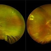
 Eales Disease
Apr 26 2013 by Howard Schatz, MD
25-year-old white female, Eales cyclitis, 2 months post vit membrantory R/D surgery; 20/70; 20/20.
Condition/keywords: Eales disease
-
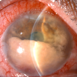
 Fuchs' Heterochromic iridocyclitis
Mar 5 2021 by Niloofar Piri, MD
Heterochromia associated with Fuchs' heterochromic iridocyclitis in the left eye of a 63-year-old patient. Notice the green color of the left iris resulting from diffuse iris atrophy.
Photographer: Douglas Snyder, MD. St. Louis University
Condition/keywords: Fuchs, Fuchs' heterochromic cyclitis, heterochromia
-
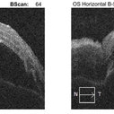
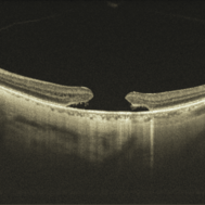 Macular Hole
Jan 3 2022 by Thirumalesh Mochi Basavaraj, MD
Swept source OCT of a 65-year-old patient with posterior hyaloid separation and a large macular hole with undermined edges.
Photographer: Puttaswamy Narayana Nethralaya Bangalore
Imaging device: Topcon Dri Triton
Condition/keywords: large macular hole, posterior cyclitis hyaloid separation, swept source OCT
-
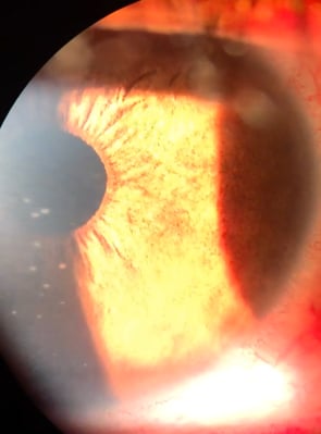 Russell bodies Observed in a Patient with Fuchs' Heterochromic Iridocyclitis
Nov 1 2016 by PAVEL FLORES-MORENO
Anterior chamber shows in iris surface: small, refractile iris crystals.
Photographer: Pavel Flores-Moreno
Imaging device: Anterior chamber camera
Condition/keywords: Fuchs, Russell bodies
-
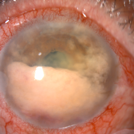
 Secondary intraocular lymphoma
Apr 11 2022 by Aniruddha K Agarwal, MD
A 65 year-old male underlying nasopharygeal non-Hodgkin’s lymhoma presented with pseudohypopyon and infiltration of iris from tumor on his right eye. Aqueous tap showed atypical lymphocytes.
Photographer: Kessara Pathanapitoon MD, PhD Department of Ophthalmology, Faculty of Medicine Chiang Mai University, Chiang Mai, Thailand
Condition/keywords: lymphoma, masquerade syndrome, secondary iridocyclitis (noninfectious)
-
 Secondary intraocular lymphoma
Apr 11 2022 by Aniruddha K Agarwal, MD
A 65-year-old male underlying nasopharyngeal non-Hodgkin’s lymphoma presented with pseudo-hypopyon and infiltration of iris from tumor on his right eye. Aqueous tap showed atypical lymphocytes.
Photographer: Kessara Pathanapitoon MD, PhD Department of Ophthalmology, Faculty of Medicine Chiang Mai University, Chiang Mai, Thailand
Condition/keywords: lymphoma, masquerade syndrome, secondary iridocyclitis (noninfectious)
-
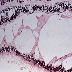
 Slide 2-25
Feb 19 2019 by Lancaster Course in Ophthalmology
Mononuclear cells in the iris and ciliary body, in a herpes simplex iridocyclitis.
Condition/keywords: ciliary, Herpes, iris, mononuclear
-
 Slide 2-35
Feb 19 2019 by Lancaster Course in Ophthalmology
Atrophic iris in Fuch's heterochromic iridocyclitis. Mild plasma cell and lymphocytic infiltrate is present, as well as small vessels in the anterior border layer.
Condition/keywords: Fuchs' heterochromic cyclitis, iris atrophy, lymphocytes
-
 Slide 9-53
Feb 26 2019 by Lancaster Course in Ophthalmology
Macular cystic degeneration and early retinoschsis in eye with chronic iridocyclitis.
Condition/keywords: cystoid macular degeneration, iridocyclitis, retinoschisis
-
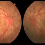 Vogt-Koyanagi-Harada
Jun 11 2016 by John S. King, MD
Initial Presentation 20/200 OU O/W healthy AAF 30-year-old c one month poor vision, occasional HAs and tinnitus; no poliosis (hx of HA eval c neg mri; hx of iridocyclitis tx)
Condition/keywords: Vogt-Koyanagi-Harada
-
 Vogt-Koyanagi-Harada
Jun 11 2016 by John S. King, MD
Initial Presentation 20/200 OU O/W healthy AAF 30-year-old c one month poor vision, occasional HAs and tinnitus; no poliosis (hx of HA eval c neg mri; hx of iridocyclitis tx)
Condition/keywords: Vogt-Koyanagi-Harada
-
 Vogt-Koyanagi-Harada
Jun 11 2016 by John S. King, MD
Initial Presentation 20/200 OU O/W healthy AAF 30-year-old c one month poor vision, occasional HAs and tinnitus; no poliosis (hx of HA eval c neg mri; hx of iridocyclitis tx)
Condition/keywords: Vogt-Koyanagi-Harada
 Loading…