Search results (236 results)
-
 Acanthamoeba Of Cornea
Jul 29 2013 by H. Michael Lambert, MD
Acanthamoeba of cornea.
Condition/keywords: acanthamoeba , cornea
-
 Applinator Prism Alcohol Burn on Cornea.
Jul 11 2013 by Jason S. Calhoun
Patient who was applinated for IOP check with applinator prism, produced a burn from the tip of the prism after it was cleaned with alcohol. Fluoresce staining shows a ring burn on the epithelium.
Photographer: Jason S. Calhoun, Department of Ophthalmology, Mayo Clinic Jacksonville, Florida
Condition/keywords: cornea
-
 Congenital Syphilis
Feb 20 2015 by H. Michael Lambert, MD
Interstitial keratitis in syphilis.
Condition/keywords: congenital, cornea, interstitial and deep keratitis, syphilis
-
 Congenital Syphilis
Feb 20 2015 by H. Michael Lambert, MD
Interstitial keratitis in syphilis.
Condition/keywords: congenital, cornea, interstitial and deep keratitis, syphilis
-
 Congenital Syphilis
Feb 20 2015 by H. Michael Lambert, MD
Interstitial keratitis in syphilis.
Condition/keywords: congenital, cornea, interstitial and deep keratitis, syphilis
-
 Cornea
Feb 23 2018 by JEFFERSON R SOUSA, Tecg.º (Biomedical Systems Technology)
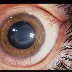
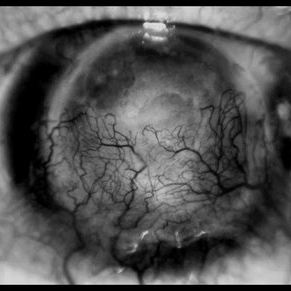
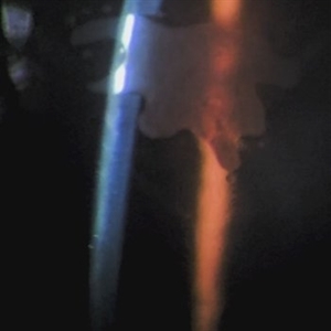
64-year-old, patient with vision loss more than 10 years after having suffered blunt trauma with ocular perforation.
Photographer: JEFFERSON R SOUSA - Study Center and Ophthalmological Research Dr. Andre M V Gomes, Institute Dr. Suel Abujamra São Paulo-Brazil
Imaging device: Topcon TRC-50 DX, Imaginet 5.0, angle de 20 graus. Flash 36.
Condition/keywords: 20 degrees, central opacity of cornea, corneal edema, neovascularization (NV)
-
 CORNEA
Feb 23 2018 by JEFFERSON R SOUSA, Tecg.º (Biomedical Systems Technology)
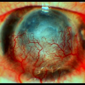
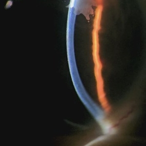
64-year-old patient, with vision loss more than 10 years after having suffered blunt trauma with ocular perforation.
Photographer: JEFFERSON R SOUSA - Study Center and Ophthalmological Research Dr. Andre M V Gomes, Institute Dr. Suel Abujamra São Paulo-Brazil
Imaging device: Topcon TRC-50 DX, Imaginet 5.0, angle de 20 graus. Flash 36.
Condition/keywords: 20 degrees, central opacity of cornea, corneal edema, neovascularization (NV)
-
---thumb.jpg/image-square;max$300,300.ImageHandler) Cornea / Toxicity Propine
Cornea / Toxicity Propine
-
---thumb.jpg/image-square;max$300,300.ImageHandler) Cornea / Toxicity Propine
Cornea / Toxicity Propine
-
 Cornea Transplant
Apr 27 2018 by Giselle DeOliveira
Cornea transplant on adult female.
Photographer: Giselle DeOliveira, University of Miami, Bascom Palmer Eye Institute
Condition/keywords: cornea, transplant
-
 Corneal Abnormal Blood Vessels
Jul 14 2013 by Jason S. Calhoun
Corneal neovascularization, abnormal blood vessels growing on the epithelium.
Photographer: Jason S. Calhoun, Department of Ophthalmology, Mayo Clinic Jacksonville, Florida
Imaging device: TOPCON D-90 SL NIKON CAMERA
Condition/keywords: cornea
-
 Corneal Abrasion
Jul 13 2013 by Jason S. Calhoun
Patient comes in with severe eye pain, blurred vision and light sensitivity.
Photographer: Jason S. Calhoun, Department of Ophthalmology, Mayo Clinic Jacksonville, Florida
Imaging device: TOPCON D-90 SL NIKON CAMERA
Condition/keywords: cornea, epithelial changes
-
 Corneal Ring Fundus Optos Photo
May 18 2020 by Catalina Montoya, MD
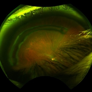
Temporal Fundus photograph right eye. Optos. No lesions in retina. Corneal Ring observed over fundus photograph. Patient with keratoconus.
Photographer: Catalina Montoya, Intermédica 1313. Medellín, Colombia
Imaging device: Optos
Condition/keywords: cornea
-
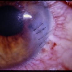 Corneal Wound - Sutured
-
 DSEK
DSEK
Jul 14 2013 by Jason S. Calhoun
Descemet's stripping endothelial keratoplasty, air bubble in anterior chamber.
Photographer: Jason S. Calhoun, Department of Ophthalmology, Mayo Clinic Jacksonville, Florida
Imaging device: TOPCON D-90 SL NIKON CAMERA
Condition/keywords: cornea
-
---thumb.JPG/image-square;max$300,300.ImageHandler) DSEK
DSEK
Jul 14 2013 by Jason S. Calhoun
Descemet's stripping endothelial keratoplasty, air bubble in anterior chamber.
Photographer: Jason S. Calhoun, Department of Ophthalmology, Mayo Clinic Jacksonville, Florida
Imaging device: TOPCON D-90 SL NIKON CAMERA
Condition/keywords: cornea
-
 IG
IG
May 17 2013 by Howard Schatz, MD
IG, 20/30; 20/100, adhesion, iris cornea.
Condition/keywords: adhesions of iris, cornea, iris
-
 IG
IG
May 17 2013 by Howard Schatz, MD
71-year-old white female, severe IG vessel on cornea.
Condition/keywords: cornea, IG
-
 Ink Blot Epithelial Ingrowth Post-LASIK Refractive Surgery
Jun 29 2024 by Luai Abu-Ismail, MD
Anterior segment photo of a 45-year-old female patient presented 12-year post-LASIK surgery.
Photographer: Dr. Luai Abu-Ismail, Ophthalmology Department, Islamic Hospital.
Imaging device: Slit lamp biomicroscope photo taken by Smart phone camera.
Condition/keywords: complication, cornea, corneal scars and opacities, epithelial ingrowth, LASIK, LASIK FLAP, refractive surgery
-
 Ink Blot Epithelial Ingrowth Post-LASIK Refractive Surgery
Jun 29 2024 by Luai Abu-Ismail, MD
Anterior segment photo of a 45-year-old female patient presented 12-year post-LASIK surgery.
Photographer: Dr. Luai Abu-Ismail, Ophthalmology Department, Islamic Hospital.
Imaging device: Slit lamp biomicroscope photo taken by Smart phone camera.
Condition/keywords: cornea, corneal scars and opacities, flap, LASIK
 Loading…