Search results (236 results)
-
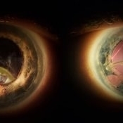
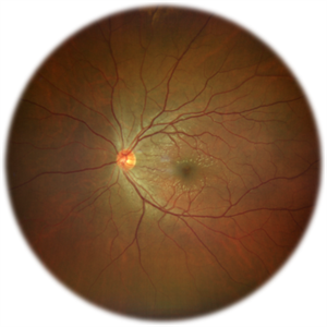 Commotio Retinae
Jun 10 2025 by CUI YUELING
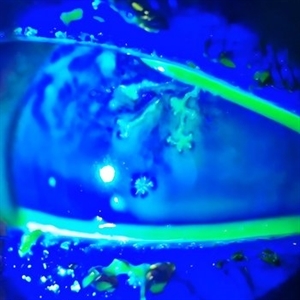
The patient presented 2 hours after sustaining a left eye injury caused by a stick. Visual acuity in the left eye was 0.2 without improvement upon correction, and intraocular pressure measured 15 mmHg. Examination of the anterior segment revealed ciliary conjunctival injection accompanied by patchy subconjunctival hemorrhage. The corneal surface remained smooth, and the anterior chamber was deep with hyphema characterized by blood-tinged aqueous humor predominantly settled inferiorly. The pupil was slightly irregular, approximately 3 mm in diameter, with a superotemporal notch; pupillary light reflex was intact. The lens appeared clear. Fundus examination showed well-defined optic disc margins with normal coloration and a cup-to-disc ratio of 0.2. Retinal arteries and veins were normally distributed with an artery-to-vein ratio of 2:3. At the posterior pole, the foveal reflex exhibited concentric ripple-like changes centered on the fovea, accompanied by localized pigment attenuation and reduced reflex intensity. Irregular reflectivity was noted in the superotemporal and inferotemporal nerve fiber layers.
Photographer: Yueling Cui
Imaging device: Zeiss Clarus 500
Condition/keywords: commotio retinae
-
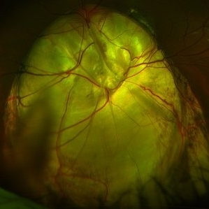
 Vitreous Bands
Apr 28 2025 by Gustavo Uriel Fonseca Aguirre
This B-mode transversal ultrasound scan shows condensed vitreous bands with anterior traction toward the cornea, accompanied by vitreous cellularity, in a patient with corneal perforation secondary to bacterial keratitis. The findings indicate severe intraocular inflammation with potential vitreous involvement.
Photographer: Gustavo U. Fonseca Aguirre, Hospital Conde de Valenciana, Ciudad de México
Condition/keywords: keratitis, vitreous bands
-
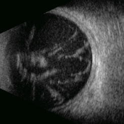
Eye of the Hurricane
Apr 9 2025 by Gustavo Uriel Fonseca Aguirre
Ultrasound biomicroscopy of a post-operative eye (status post trabeculectomy and phacoemulsification) reveals a patent ostium on the right side, along with an intraocular lens in position. A hyphema is observed displaying small convection currents, creating a circular motion pattern due to the temperature gradient between the iris and cornea. Notably, the blood flow can be seen circulating toward the trabeculectomy site.
Condition/keywords: hyphema, trabeculectomy
-
 Eye of the Hurricane
Apr 8 2025 by Gustavo Uriel Fonseca Aguirre
Ultrasound biomicroscopy of a post-operative eye (status post trabeculectomy and phacoemulsification) reveals a patent ostium on the right side, along with an intraocular lens in position. A hyphema is observed displaying small convection currents, creating a circular motion pattern due to the temperature gradient between the iris and cornea. Notably, the blood flow can be seen circulating toward the trabeculectomy site.
Photographer: Gustavo U. Fonseca Aguirre, Hospital Conde de Valenciana, Ciudad de México
Condition/keywords: Hyphema, trabeculectomy
-
 Ozurdex in AC
Apr 1 2025 by Korey Starkey
90-year-old patient with an Ozurdex implant that migrated into the AC and with the cornea decompensating. Patient recommended for urgent surgery to remove implant. Vision OD at this visit was CF @ 2ft, most recent visit vision is 20/400, PH 20/25.
Photographer: Korey Starkey
Imaging device: Topcon
Condition/keywords: anterior chamber, corneal decompensation, external, external photography, Ozurdex implant, Topcon
-
 Retisert Implant Migration Into the Anterior Chamber
Feb 11 2025 by Niloofar Piri, MD
Slit Lamp photograph demonstrating spontaneous dislocation and migration of old Retisert implant into the anterior chamber inferiorly with secondary corneal decompensation. Please notice that patient is aphakic. Implant was removed surgically.
Photographer: Hossein Asghari MD, Saint Louis University
Condition/keywords: Corticosteroid implant, implant migration, Retisert
-
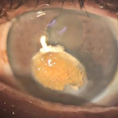
 Bullous Keratopathy
Jan 4 2025 by Mosab Salah
Corneal Slit photograph of an 84-year-old man underwent uneventful cataract surgery 1 year ago elsewhere, with a multiple fluid filled Bullae, not responding on conservative management and planned for KP.
Photographer: Abu-Ismail, Luai MD, The Islamic Hospital, Amman, Jordan
Imaging device: smartphone photography through SLB
Condition/keywords: bullous keratopathy, corneal edema
-
 Rosai-Dorfman Disease
Dec 4 2024 by Virginia Gebhart
72 year old female with temporal limbal lesion that extends onto the cornea from 10:00 - 8:00 encroaching on visual axis. Possible lymphomatous process. Will refer to Emory.
Photographer: Dr Chris Bergstrom MD, OD
Condition/keywords: corneal scars and opacities, Rosai-Dorfman Disease, Subconjunctival mass
-
 Expulsion of Retina
Oct 23 2024 by Gustavo Uriel Fonseca Aguirre
Male patient with a history of penetrating keratopathy presents due to blunt ocular trauma. A disruption of the continuity at the interface between the donor and recipient corneas is observed, with expulsion of the lens and retina. Vision is limited to light perception.
Photographer: Lizeth Jiménez Santana, Fundación Hospital Nuestra Señora de la Luz, Ciudad de México
Condition/keywords: ocular trauma, penetrating keratoplasty
-
 Spheroidal Degeneration
Sep 28 2024 by DR Rohit Gupta
Slit lamp photograph of a 68 year-old male patient presented with diminution of vision and foreign body sensation. On examination brown cataract with yellowish globular degeneration seen on cornea.
Photographer: Dr Rohit gupta
Imaging device: Samsung S21
Condition/keywords: Spheroidal degeneration
-
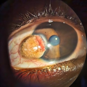 Limbal Dermoid
Sep 25 2024 by DR Rohit Gupta
Slit lamp photograph of a 22 year-old female presenting with a swelling over cornea which on examination appears be to be Limbal dermoid.
Photographer: Dr Rohit gupta
Imaging device: Samsung S21
Condition/keywords: dermoid, limbus, lipodermoid
-
 Herpes Simplex
Sep 24 2024 by DR Rohit Gupta
Slit lamp photograph of a 32 year-old male presented with redness, photophobia, and pain in left eye.
Photographer: Dr Rohit gupta
Imaging device: Samsung S21
Condition/keywords: corneal ulcer, Herpes, herpes dendrite, Herpes simplex infection
-
 Herpetic Corneal Ulcer
Sep 24 2024 by DR Rohit Gupta
Slit lamp photograph of 32 year old male presented with herpetic corneal ulcer on staining with fluorescein dye under cobalt blue filted dendrits can be seen.
Photographer: Dr Rohit gupta
Imaging device: Samsung S21
Condition/keywords: corneal ulcer, dendritic keratitis, herpes dendrite, Herpes simplex infection, Herpes zoster, staining
-
 Ink Blot Epithelial Ingrowth Post-LASIK Refractive Surgery
Jun 29 2024 by Luai Abu-Ismail, MD
Anterior segment photo of a 45-year-old female patient presented 12-year post-LASIK surgery.
Photographer: Dr. Luai Abu-Ismail, Ophthalmology Department, Islamic Hospital.
Imaging device: Slit lamp biomicroscope photo taken by Smart phone camera.
Condition/keywords: cornea, corneal scars and opacities, flap, LASIK
-
 Ink Blot Epithelial Ingrowth Post-LASIK Refractive Surgery
Jun 29 2024 by Luai Abu-Ismail, MD
Anterior segment photo of a 45-year-old female patient presented 12-year post-LASIK surgery.
Photographer: Dr. Luai Abu-Ismail, Ophthalmology Department, Islamic Hospital.
Imaging device: Slit lamp biomicroscope photo taken by Smart phone camera.
Condition/keywords: complication, cornea, corneal scars and opacities, epithelial ingrowth, LASIK, LASIK FLAP, refractive surgery
-
 Dislocated IOL
Jun 4 2024 by Marlee Curnutt
Slit lamp photo of a 64 year old woman presenting with worsening vision and depth perception after trauma induced by a dog, which dislocated her IOL. The patient's IOL haptic was seen in the AC, and almost abutting cornea. Patient's vision upon presentation was DCC CF@1 feet. Patient was counseled and underwent an IOL exchange.
Photographer: Marlee Curnutt, COA
Imaging device: Galaxy A42
Condition/keywords: dislocated intraocular lens (IOL), haptic, IOL, right eye, slit lamp photo, slit lamp photography, trauma
-
 Keratoconus - Corneal Scar
May 3 2023 by Paula Lavigne
Keratoconus - Corneal Scar
Photographer: Paula Lavigne, Obras Sociais Irmã Dulce
Condition/keywords: corneal ectasia, corneal scars and opacities, keratoconus
-
 Fundus Coloboma
Feb 22 2023 by Zach Seim
An ultra-widefield fundus image of a 25 year old male with Fundus Coloboma, as well as Iris Coloboma affecting both eyes. Patient's vision at the time of the image was 20/100-2. Discussed genetic testing as patient reports that he has a child with coloboma and patient agrees. There is a possibility of this finding being syndromic given cornea has small WTW and possibly microphthalmia. The patient has old tractional exudation at edge (abutting fovea). Recommended observation without treatment.
Photographer: Zach Seim
Imaging device: Optos California
Condition/keywords: coloboma, coloboma of optic disc, fundus photograph, Optos, scanning laser ophthalmoscope, ultra-wide field imaging
-
 Iris Coloboma
Feb 22 2023 by Zach Seim
An external image of a 25 year old male with Iris Coloboma, as well as Fundus Coloboma affecting both eyes. Patient's vision at the time of the image was 20/80. Discussed genetic testing as patient reports that he has a child with coloboma and patient agrees. There is a possibility of this finding being syndromic given cornea has small WTW and possibly microphthalmia. Recommended observation without treatment.
Photographer: Zach Seim
Imaging device: Topcon 50DX
Condition/keywords: coloboma, iris, left eye, Topcon
-
 Posterior Scleral Laceration
May 24 2022 by Ahmad B. Tarabishy, MD
A 49 year old male was referred from the ER following an injury to his right medial eyelid with a sharp metal tip. He had brief pain at the time. No new floaters, flashes, or blurred vision. Intraocular pressure was 18 OS. Examination showed a full thickness laceration of the nasal posterior globe with adjacent hemorrhage. Prophylactic laser coagulation was performed. Examination 2 weeks later shows maturing laser scars and no complications related to the scleral laceration. The patient reports no new vision changes.
Photographer: Angelo Rico MD, Retina Specialists of Tampa
Imaging device: Optos
Condition/keywords: corneal laceration, globe perforation
-
 Acute Anterior Uveitis
Apr 14 2022 by Divya Jain
Anterior Segment Slit Lamp photograph of a 33 year old woman with first episode of acute granulomatous anterior uveitis showing circumcorneal congestion, mutton fat KP'S, 3+ cells, 2+ flare and Koeppe's nodules at pupillary margin.
Photographer: Divya Jain
Condition/keywords: acute anterior uveitis
-
 The Effects of Blunt Trauma
Feb 27 2022 by Jesus Lozano, MD
Axial Head Ct. 60 year old man with a history of blunt trauma and lost of vision after the event. VA HM. Iop 25mmhg. Cornea clear. Complete hyphema. BMode US: diffuse Vitreous Hemorrhage with a Dislocated Lens. Retina attached.
Photographer: Dr. Jesus Lozano. Retina Specialist. Hillel Yaffe Medical Center,Israel.
Imaging device: Axial Head CT
Condition/keywords: blunt trauma, hyphema, lens dislocation
 Loading…