Search results (236 results)
-
 Cornea Transplant
Apr 27 2018 by Giselle DeOliveira
Cornea transplant on adult female.
Photographer: Giselle DeOliveira, University of Miami, Bascom Palmer Eye Institute
Condition/keywords: cornea, transplant
-
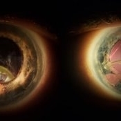
Eye of the Hurricane
Apr 9 2025 by Gustavo Uriel Fonseca Aguirre
Ultrasound biomicroscopy of a post-operative eye (status post trabeculectomy and phacoemulsification) reveals a patent ostium on the right side, along with an intraocular lens in position. A hyphema is observed displaying small convection currents, creating a circular motion pattern due to the temperature gradient between the iris and cornea. Notably, the blood flow can be seen circulating toward the trabeculectomy site.
Condition/keywords: hyphema, trabeculectomy
-
 Eye of the Hurricane
Apr 8 2025 by Gustavo Uriel Fonseca Aguirre
Ultrasound biomicroscopy of a post-operative eye (status post trabeculectomy and phacoemulsification) reveals a patent ostium on the right side, along with an intraocular lens in position. A hyphema is observed displaying small convection currents, creating a circular motion pattern due to the temperature gradient between the iris and cornea. Notably, the blood flow can be seen circulating toward the trabeculectomy site.
Photographer: Gustavo U. Fonseca Aguirre, Hospital Conde de Valenciana, Ciudad de México
Condition/keywords: Hyphema, trabeculectomy
-
 4 Point Scleral Fixation Akreos AO60 With Gore Tex Suture
May 21 2021 by Jesus Lozano, MD
Anterior segment photo of a 54-year-old man after 4 point scleral fixation Akreos AO60 with Gore Tex suture plus PPV who had a severe traumatic iris defect and was aphakic after ocular trauma.
Photographer: Luigi Zinn, Hadassah Medical Center, Jerusalem.
Condition/keywords: aphakia, cornea rupture, lens, penetrating trauma
-
 Applinator Prism Alcohol Burn on Cornea.
Jul 11 2013 by Jason S. Calhoun
Patient who was applinated for IOP check with applinator prism, produced a burn from the tip of the prism after it was cleaned with alcohol. Fluoresce staining shows a ring burn on the epithelium.
Photographer: Jason S. Calhoun, Department of Ophthalmology, Mayo Clinic Jacksonville, Florida
Condition/keywords: cornea
-
 Chorioretinal Coloboma with Retinal Detachment
Dec 5 2020 by Niloofar Piri, MD
14-year-old female with 1q21.1 microdeletion syndrome and behavioral, intellectual, and systemic abnormalities, including congenital microcornea, iris coloboma, and chorioretinal and optic nerve coloboma presented with decreased vision. Right eye fundus taken with RetCam shows coloboma with retinal detachment. (Left eye showed white cataract with funnel RD on B-scan).
Photographer: Niloofar Piri MD, Douglas Snyder MD
Condition/keywords: chorioretinal coloboma, optic nerve coloboma
-
---thumb.jpg/image-square;max$300,300.ImageHandler) Cornea / Toxicity Propine
Cornea / Toxicity Propine
-
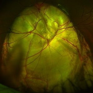
 Corneal Abnormal Blood Vessels
Jul 14 2013 by Jason S. Calhoun
Corneal neovascularization, abnormal blood vessels growing on the epithelium.
Photographer: Jason S. Calhoun, Department of Ophthalmology, Mayo Clinic Jacksonville, Florida
Imaging device: TOPCON D-90 SL NIKON CAMERA
Condition/keywords: cornea
-
 Dislocated IOL
Jun 4 2024 by Marlee Curnutt
Slit lamp photo of a 64 year old woman presenting with worsening vision and depth perception after trauma induced by a dog, which dislocated her IOL. The patient's IOL haptic was seen in the AC, and almost abutting cornea. Patient's vision upon presentation was DCC CF@1 feet. Patient was counseled and underwent an IOL exchange.
Photographer: Marlee Curnutt, COA
Imaging device: Galaxy A42
Condition/keywords: dislocated intraocular lens (IOL), haptic, IOL, right eye, slit lamp photo, slit lamp photography, trauma
-
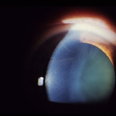
---thumb.JPG/image-square;max$300,300.ImageHandler) DSEK
DSEK
Jul 14 2013 by Jason S. Calhoun
Descemet's stripping endothelial keratoplasty, air bubble in anterior chamber.
Photographer: Jason S. Calhoun, Department of Ophthalmology, Mayo Clinic Jacksonville, Florida
Imaging device: TOPCON D-90 SL NIKON CAMERA
Condition/keywords: cornea
-
 Endophthalmitis
Apr 9 2014 by Aleksandra V. Rachitskaya, MD, FASRS
Slit lamp photo of a patient with endophthalmitis after cataract surgery. An infectious infiltrate is noted next to the clear corneal incision.
Photographer: Bascom Palmer Eye Institute
Condition/keywords: cataract surgery, endophthalmitis
-
 Expulsion of Retina
Oct 23 2024 by Gustavo Uriel Fonseca Aguirre
Male patient with a history of penetrating keratopathy presents due to blunt ocular trauma. A disruption of the continuity at the interface between the donor and recipient corneas is observed, with expulsion of the lens and retina. Vision is limited to light perception.
Photographer: Lizeth Jiménez Santana, Fundación Hospital Nuestra Señora de la Luz, Ciudad de México
Condition/keywords: ocular trauma, penetrating keratoplasty
-
 Fabry's Disease Carrier
Jun 4 2014 by Henry J. Kaplan, MD
A carrier of Fabry's disease who demonstrates cornea verticillata.
Condition/keywords: cornea verticillata, Fabry disease
-
 Fundus Coloboma
Feb 22 2023 by Zach Seim
An ultra-widefield fundus image of a 25 year old male with Fundus Coloboma, as well as Iris Coloboma affecting both eyes. Patient's vision at the time of the image was 20/100-2. Discussed genetic testing as patient reports that he has a child with coloboma and patient agrees. There is a possibility of this finding being syndromic given cornea has small WTW and possibly microphthalmia. The patient has old tractional exudation at edge (abutting fovea). Recommended observation without treatment.
Photographer: Zach Seim
Imaging device: Optos California
Condition/keywords: coloboma, coloboma of optic disc, fundus photograph, Optos, scanning laser ophthalmoscope, ultra-wide field imaging
-
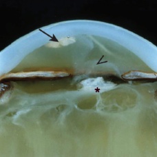 Granulomatous Uveitis
May 18 2020 by McGill University Health Centre
Granulomatous uveitis is found in many inflammatory diseases, and is generally characterized by a predominant histiocytic infiltrate forming a “wall” (granuloma) around a pathogen or foreign body. This is an example of granulomatous uveitis. The eye is aphakic; the uveal track is thickened; and a granuloma is present and attached to the endothelium of the cornea (arrow). The anterior chamber is filled with a hazy material (arrowhead). The vitreous is fibrotic and tractional bands are also present (*).
Condition/keywords: granulomatous uveitis
-
 Ozurdex in AC
Apr 1 2025 by Korey Starkey
90-year-old patient with an Ozurdex implant that migrated into the AC and with the cornea decompensating. Patient recommended for urgent surgery to remove implant. Vision OD at this visit was CF @ 2ft, most recent visit vision is 20/400, PH 20/25.
Photographer: Korey Starkey
Imaging device: Topcon
Condition/keywords: anterior chamber, corneal decompensation, external, external photography, Ozurdex implant, Topcon
-
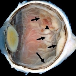 Primary Retinal and Vitreous Large B-Cell Lymphomas
May 18 2020 by McGill University Health Centre
These tumors are associated with intracranial nervous system lymphomas. The image shows an enucleation specimen with a multifocal, necrotic, and hemorrhagic whitish retinal tumor (arrow). Note the thickened, opaque cornea; the cataractous lens; the diffuse, flat retinal detachment; and the retinal hemorrhages overlying the tumor (arrowhead).
Condition/keywords: large b cell lymphoma of the retina, lymphoma
-
 Retisert Implant Migration Into the Anterior Chamber
Feb 11 2025 by Niloofar Piri, MD
Slit Lamp photograph demonstrating spontaneous dislocation and migration of old Retisert implant into the anterior chamber inferiorly with secondary corneal decompensation. Please notice that patient is aphakic. Implant was removed surgically.
Photographer: Hossein Asghari MD, Saint Louis University
Condition/keywords: Corticosteroid implant, implant migration, Retisert
-
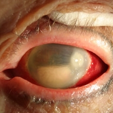
 Subconjuntival IOL After Blunt Trauma
Jun 27 2018 by Gabriel Costa Andrade, PhD
A 73-year-old male patient was referred to our ophthalmic emergency department with complaints of redness, pain, and diminution of vision in his left eye, after fall from height. The patient underwent small incision cataract surgery with polymethylmethacrylate (PMMA) IOL implantation in both the eyes 15 years back through superior sclerocorneal incision under local anesthesia. His best-corrected visual acuity was perception of light in the left eye. Ophthalmic examination using slit lamp biomicroscopy of the left eye revealed diffuse subconjunctival hemorrhage with no conjunctival laceration and inferior bulbar conjunctiva showed traumatic pseudophacocele with a sign “golden half ring,” suggesting the presence of PCIOL in subconjunctival space.There was total hyphema obscuring the view of rest of the ocular structures in his left eye.
Photographer: Gabriel Andrade, RETINA CLINIC, São Paulo, BRAZIL
Condition/keywords: dislocated intraocular lens (IOL), trauma
-
 Iris Coloboma
Feb 22 2023 by Zach Seim
An external image of a 25 year old male with Iris Coloboma, as well as Fundus Coloboma affecting both eyes. Patient's vision at the time of the image was 20/80. Discussed genetic testing as patient reports that he has a child with coloboma and patient agrees. There is a possibility of this finding being syndromic given cornea has small WTW and possibly microphthalmia. Recommended observation without treatment.
Photographer: Zach Seim
Imaging device: Topcon 50DX
Condition/keywords: coloboma, iris, left eye, Topcon
-
 Silicone Oil Retention Sutures
May 13 2019 by Andrew W. Eller, MD
48-year-old male sustained trauma including retinal detachment. Due to irregular pupil, 10-0 Prolene silicone oil retention sutures were placed to protect the cornea from keratopathy.
Photographer: Gary Vagstad, UPMC Eye Center, University of Pittsburgh
Condition/keywords: iris, silicone oil, trauma
-
 Retinal Detachment Repair With Silicone Oil and Scleral Buckle, Fourteen Years Later, With Visual Acuity of 20/25
Sep 12 2016 by Timothy S Fuller, MD
65-year-old woman s/p scleral buckle 14 years ago. Two weeks later, the retina re-detached, and vitrectomy, laser, and silicone oil procedure was performed. Patient remains 20/25 with correction fourteen years later. The cornea is clear, there is no oil emulsification, and there is a stable, moderately inferiorly subluxated PCIOL (as it was prior to RD surgery). IOP is 17 on Cosopt BID.
Photographer: Nicholas Hesse, Texas Retina Associates
Imaging device: Optos
Condition/keywords: laser, scleral buckle, silicone oil
-
 CORNEA
Feb 23 2018 by JEFFERSON R SOUSA, Tecg.º (Biomedical Systems Technology)
64-year-old patient, with vision loss more than 10 years after having suffered blunt trauma with ocular perforation.
Photographer: JEFFERSON R SOUSA - Study Center and Ophthalmological Research Dr. Andre M V Gomes, Institute Dr. Suel Abujamra São Paulo-Brazil
Imaging device: Topcon TRC-50 DX, Imaginet 5.0, angle de 20 graus. Flash 36.
Condition/keywords: 20 degrees, central opacity of cornea, corneal edema, neovascularization (NV)
-
 24 Hours Post Scleral Wound Closure+ Scleral Buckle+25 g Vitrectomy+Silicon Oil
Jan 23 2015 by Carlos Quezada-Ruiz, MD, FASRS
24 hours post op fundus photograph of a 43-year-old man who had perforating injury to the right eye with a small piece of plastic while he was hammering. OD LP, subconjunctival hemorrhage, clear cornea, hyphema, irido and ciclodyalisis as well as a luxated lens with traumatic cataract and a dense vitreous hemorrhage. B-US showed rhegmatogenous retinal detachment with a tear and a big inferior hemorrhagic choroidal detachment. 360 peritomy revealed 2-entry scleral wounds were found in zone II (M V and M VI) and closure was performed. 25 G PPV was performed with the infusion canal placed in the AC through the limbus. Lensectomy and removal of a dense recent vitreous hemorrhage revealed a white detached retina with an exit wound through the temporal inferior segment of the optic nerve with a nasal GRT and sub retinal hemorrhage as well as temporal inferior choroidal, PVD was induced and PFOs helped stabilizing the retina while vitrectomy and sub-retinal hemorrhage was removed through the GRT. Fluid air exchange was made and 360 endolaser over the buckle indentation was done and silicon oil was used as endotamponade. This picture was taken 24 hrs after the surgery.
Photographer: Lilibeth Rodriguez, Instituto de la Visión. Torreon, Mexico.
Condition/keywords: central retinal artery occlusion (CRAO), giant retinal tear, trauma
 Loading…