 Initializing download.
-
 By Niloofar Piri, MD
SSM Health Group, St Louis University
Co-author(s): Henry J Kaplan MD, University of Louisville - Uploaded on Mar 13, 2015.
- Last modified by Chayal Patel on Jun 3, 2015.
- Image of the week
-
May 31, 2015
View all images of the week - Rating
- Appears in
- Miscellaneous
- Condition/keywords
- HIV, CMV retinitis
- Photographer
- Angela Anderson
- Imaging device
- Fundus camera
- Description
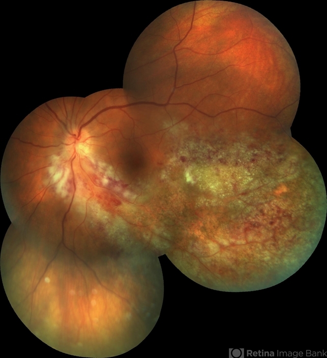
- Fundus photograph of the left eye of a 40-year-old Caucasian female with history of positive HIV test for 23 years. She has been off HAART therapy for the past 2 years and presented with decreased vision OS and upper visual field defect. On examination, she had trace cells in anterior vitreous , hemorrhagic retinitis which starts around the optic nerve and extending to inferotemporal arcade with secondary inferotemporal BRVO; in temporal periphery , she had granular pattern of CMV retinitis which is a manifestation of outer retina involvement.


---thumb.jpg/image-square;max$79,0.ImageHandler "CMV Retinitis")
---thumb.jpg/image-square;max$79,0.ImageHandler "CMV Retinitis")
---thumb.jpg/image-square;max$79,0.ImageHandler "CMV Retinitis")
---thumb.jpg/image-square;max$79,0.ImageHandler "CMV Retinitis")


