Search results (274 results)
-
 Meridional Fold
Nov 9 2012 by Norman Byer
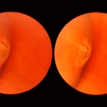
This is the same lesion as in the previous photograph. With the scleral indentation placed more posterior, we now can see that the fold ends over a small collection of subretinal fluid and that there is a very tiny retinal hole just below the posterior end of the retinal fold.
Condition/keywords: peripheral cystoid degeneration, retinal fold, retinal hole, scleral indentation, subretinal fluid
-
 White Retinal Tuft
Nov 9 2012 by Norman Byer
After six years, the previous lesion looked like this. The former flap has been completely avulsed and is now a free operculum. The white zone around the tear represents the small area of detachment and subretinal fluid. It is still asymptomatic and does not require treatment.
Condition/keywords: does not require treatment, free operculum, operculated retinal hole, subretinal fluid, white retinal tuft
-
 Lattice Degeneration
Nov 9 2012 by Norman Byer
This 16-year-old girl has lattice degeneration and also this large oval retinal hole with a surrounding narrow zone of subretinal fluid. This lesion illustrates how large the atrophic holes of lattice degeneration may be. Occasionally the hole can be as large as the initial lattice lesion and can therefore obliterate all other evidence of its true identity. This was almost true in this case, but there does remain a small whitish remnant of the original lattice lesion at the lower end of the oval hole.
Condition/keywords: lattice degeneration, retinal hole, subretinal fluid, white lattice lines
-
 Optos Giant Tear within Retinal Detachment
Apr 30 2019 by Lauren Whaley
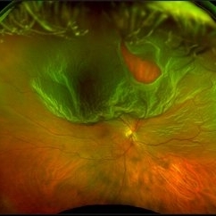
Noticed an inferior visual field defect on a patient with history of vitreous hemorrhage. Decided to take an Optos image and this is what we found. Doctor performed pneumatic retinopexy in office and patient recovering well.
Photographer: Lauren R. Whaley
Imaging device: Optos
Condition/keywords: Optos, retinal tear, subretinal fluid
-
 Choroidal Melanoma Large Amelanotic
Oct 15 2012 by Susanna S. Park, MD, PhD
68-year-old man with a large amelanotic mass and subretinal fluid in the left eye. Visual acuity was CF and extrascleral extension was suspected on MRI scan.
Photographer: Ellen Redenbo, University of California Davis Eye Center
Condition/keywords: melanoma
-
 Recurrent Central Serous Choroidopathy
Aug 21 2012 by Edwin H. Ryan, MD
EDI-OCT showing thickened choroid and subretinal fluid
Photographer: Edwin Ryan Jr. MD, VitreoRetinal Surgery, PA
Imaging device: Heidelberg Spectralis
Condition/keywords: central serous chorioretinopathy (CSCR), choroidal thickening, enhanced depth imaging
-
 Asymptomatic Lesion
Nov 9 2012 by Norman Byer
This asymptomatic lesion in a 27-year-old woman is a very interesting example of a cystic retinal tuft. Note the discrete white nubbin, which is the chief characteristic of this lesion. In this case, it is surrounded by a small area of subretinal fluid. The next slide pair will reveal the reason for this.
Condition/keywords: asymptomatic, cystic retinal tuft, subretinal fluid
-
 Chronic Macular Hole
Sep 2 2012 by Hyung-Woo Kwak, MD
A large hole with rolled everted edges, adjacent cystoid intraretinal spaces, a shallow rim of subretinal fluids.
Imaging device: Zeiss F450 plus
Condition/keywords: macular hole
-
 Central Serous Chorioretinopathy
Aug 23 2012 by Gerardo Garcia-Aguirre, MD
Fundus photograph of a 29 year-old patient showing subretinal fluid in the macula.
Photographer: Noemí Hernández, Asociación para Evitar la Ceguera en México
Imaging device: Zeiss FF4
Condition/keywords: central serous chorioretinopathy (CSCR)
-
 White Retinal Tuft
Nov 9 2012 by Norman Byer
This white retinal tuft was seen in a 20-year-old man. It is associated with an asymptomatic retinal tear posterior to the tuft and with a tiny adjacent amount of subretinal fluid. It remained just like this for six years and then underwent the change shown in the next slide pair.
Condition/keywords: asymptomatic, subretinal fluid, white retinal tuft
-
---thumb.JPG/image-square;max$300,300.ImageHandler) Retinal Pigment Epithelial Detachment With No Subretinal Fluid
Retinal Pigment Epithelial Detachment With No Subretinal Fluid
Jun 29 2013 by Jason S. Calhoun
A 38-year-old male who comes in with blurred vision in the left eye. VA is 20/30. Noticed a defect inferior of his central vision. Did an fluorescein angiogram to determine an RPE with no sub retinal fluid. Also OCT confirms. Patient was injected with Avastin.
Photographer: Jason S. Calhoun, Mayo Clinic Jacksonville, Florida
Imaging device: TOPCON TRC 50-EX
Condition/keywords: central serous retinopathy (CSR), retinal pigment epithelium (RPE) detachment
-
 OCT of Large Choroidal Nevus
Jul 9 2014 by Susanna S. Park, MD, PhD
EDI OCT imaging of a large pigmented choridal nevus showing mild elevation with no associated subretinal fluid.
Photographer: Ellen Redenbo
Condition/keywords: choroidal nevus, optical coherence tomography (OCT)
-
 Lattice Lesion
Nov 9 2012 by Norman Byer
This is the same lesion as shown in the previous case. Two retinal holes are present, and you can look through the upper hole into the dark subretinal space. This is, therefore, a true subclinical retinal detachment but it has changed only slightly in the past 13 years. About 75% of such holes in lattice lesions show a tiny adjacent zone of subretinal fluid. After the hole forms from gradual progressive thing of the retina, a tiny amount of fluid from the pocket of liquified vitreous over the lesion passes through the hole to the subretinal space
Condition/keywords: lattice degeneration, liquefied vitreous, retinal hole, subretinal fluid
-
 Macular Hole
Sep 27 2012 by Jeffrey G. Gross, MD, FASRS
Macular hole s/p 360 degree laser to fluid cuff.
Condition/keywords: macular hole, subretinal fluid
-
 Bulls eye retinopathy
Nov 20 2012 by Roy Schwartz, MD
75-YEAR-OLD FEMALE PRESENTS WITH BILATERAL GRADUAL VISUAL LOSS 6/30 Dx BE PSEUDOPHAKIA + PCO BE BULLS EYE MACULOPATHY PER FA VA IMPROVES TO 6/10 S/P YAG CAPSULOTOMY OCT - BE MACULAR SUBRETINAL FLUID NO HISTORY OF CHLOROQUINE THERAPY NO DRUSEN OR SIGNS OF AMD WORKING DIAGNOSIS - BE CHRONIC CSCR
Imaging device: Heidelberg spectralis
Condition/keywords: bull's eye maculopathy, optical coherence tomography (OCT)
-
 Scleral Indentation
Nov 9 2012 by Norman Byer
This is the same lesion with scleral indentation. You can see the small discrete preretinal hemorrhage and the sharply circumscribed area of elevated retina with subretinal fluid beneath it. No retinal break is visible, but the posterior vitreous is detached and exerting traction at this site. The area was surrounded with argon laser treatment the same day as the initial examination.
Condition/keywords: posterior vitreous detachment, preretinal hemorrhage, scleral indentation, subretinal fluid, vitreous traction
-
 Choroidal Melanoma
Feb 2 2018 by Olivia Rainey
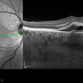
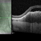
Optical coherence tomography with enhanced depth imaging of a 78-year-old female with choroidal melanoma with subretinal fluid affecting her right eye.
Photographer: Olivia Rainey
Imaging device: Heidelberg Spectralis
Condition/keywords: enhanced depth imaging, infrared image, optical coherence tomography (OCT), subretinal fluid, superior retina
-
 Choroidal Granuloma Secondary to Tuberculosis
Mar 14 2013 by Eduardo Torres-Porras, MD
OCT scan through the granuloma shows attachment of the retinal pigment epithelial-choriocapillaris layer and the neurosensory retina over the granuloma (“contact” sign), inflammatory retinal infiltrate in the deeper retinal layers and subretinal fluid.
Photographer: Eduardo Torres Porras, Laser y ultrasonido ocular de Puebla
Imaging device: Cirrus
Condition/keywords: optical coherence tomography (OCT), tubercular choroidal granuloma
-
 EDI OCT Detachment With No Subretinal Fluid
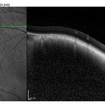
Jun 29 2013 by Jason S. Calhoun
A 38-year-old male came in with blurred vision in the left eye. VA is 20/30. Notice a defect inferior of his central vision. Did an fluorescien angiogram to determine an RPE with no subretinal fluid. Also OCT confirms. Patient was injected with Avastin.
Photographer: Jason S. Calhoun, Mayo Clinic Jacksonville, Florida
Imaging device: TOPCON TRC 50-EX/CIRRUS HD OCT
Condition/keywords: central serous retinopathy (CSR), retinal pigment epithelium (RPE) detachment
-
 Chronic Total Retinal Detachment
Oct 12 2012 by Jeffrey G. Gross, MD, FASRS
Chronic, total RD, with shifting inferior subretinal fluid.
Condition/keywords: chronic, inferior subretinal fluid
-
 Choroidal Granuloma Secondary to Tuberculosis
Mar 14 2013 by Eduardo Torres-Porras, MD
OCT scan through the granuloma shows attachment of the retinal pigment epithelial-choriocapillaris layer and the neurosensory retina over the granuloma (“contact” sign), inflammatory retinal infiltrate in the deeper retinal layers and subretinal fluid.
Photographer: Eduardo Torres Porras
Imaging device: Cirrus
Condition/keywords: optical coherence tomography (OCT), tubercular choroidal granuloma
-
 Macular Hole
Sep 27 2012 by Jeffrey G. Gross, MD, FASRS
Macular hole magnified with cuff of SRF.
Condition/keywords: cuff, macular hole, subretinal fluid
-
---thumb.JPG/image-square;max$300,300.ImageHandler) Central Serous Retinopathy with Fibrin
Central Serous Retinopathy with Fibrin
Oct 13 2012 by Edwin H. Ryan, MD
Recurrent central serous with fibrin in a 54-year-old man.
Condition/keywords: central serous chorioretinopathy (CSCR), subretinal fluid
-
 Bulls eye retinopathy OCT LE
Nov 20 2012 by Roy Schwartz, MD
75-YEAR-OLD FEMALE PRESENTS WITH BILATERAL GRADUAL VISUAL LOSS 6/30 Dx BE PSEUDOPHAKIA + PCO BE BULLS EYE MACULOPATHY PER FA VA IMPROVES TO 6/10 S/P YAG CAPSULOTOMY OCT - BE MACULAR SUBRETINAL FLUID NO HISTORY OF CHLOROQUINE THERAPY NO DRUSEN OR SIGNS OF AMD WORKING DIAGNOSIS - BE CHRONIC CSCR
Imaging device: Heidelberg spectralis
Condition/keywords: bull's eye maculopathy, chronic central serous chorioretinopathy (CSCR), optical coherence tomography (OCT)
-
 Central Serous Choroidopathy, CSR, with Foci of Leakage
Oct 9 2012 by James B. Soque, CRA, OCT-C, COA, FOPS
50 y/o WM with Central Serous Choroidopathy Left eye. VA OS cc 20/80. Topcon 3D 1000 SD OCT composite image reveals Sub RPE detachment in several locations, and subretinal fluid blister. Pin Point Registration shows leakage along horizontal line axis, SR leakage, and RPE detachments OS. Color, Early, and Late phase FA photos enclosed above. FA shows obvious ‘smoke stack’ appearance of leakage in superonasal fovea, and 3 other foci of leakage. 3D 1000 SD OCT with pin point registration image shown.
Photographer: James Soque CRA COA
Imaging device: Topcon 3D OCT 1000 System
Condition/keywords: central serous retinopathy (CSR)
 Loading…