Search results (50 results)
-
 Chronic Retinal Detachment: Features Slide 1
Oct 22 2012 by Ronald C. Gentile, MD
Chronic retinal detachments can be associated with demarcation lines (tidemarks), subretinal bands or sheets, and retinal cysts. Fundus photo of a chronic inferior retinal detachment reveals multiple demarcation lines inferior to the center of the fovea as a result of an inferior temporal dialysis.
Photographer: The New York Eye & Ear Infirmary Department of Medical Imaging
Condition/keywords: chronic retinal detachment, demarcation line
-
 Chronic Retinal Detachment
Oct 12 2012 by Jeffrey G. Gross, MD, FASRS
Chronic RD with multiple retinal cysts, B scan ultrasound.
Condition/keywords: B scan ultrasound, chronic retinal detachment, retinal cyst
-
 Chronic Retinal Detachment: Features Slide 2
Oct 22 2012 by Ronald C. Gentile, MD
Chronic retinal detachments can be associated with demarcation lines (tidemarks), subretinal bands or sheets, and retinal cysts. Fundus photo of a chronic retinal detachment reveals a branching subretinal band superior nasal to the macula with a portion extending to the inferior margin of the optic disc.
Photographer: The New York Eye & Ear Infirmary Department of Medical Imaging
Condition/keywords: chronic retinal detachment, subretinal bands
-
 Retinal Lesion
Nov 9 2012 by Norman Byer
This 30-year-old man sustained a severe blow to his brow region which resulted in a variety of injuries including hyphema and vitreous and retinal hemorrhages. This photograph shows a retinal lesion, which is either a tiny area of elevated full thickness retina or a traumatic retinal cyst.
Condition/keywords: elevated retinal lesion, retinal hemorrhage, traumatic retinal cyst
-
 Retinal Detachment
Nov 9 2012 by Norman Byer
This eye of a 25-year-old man has a retinal detachment of about six year’s duration. This photograph shows an intraretinal cyst, which is a secondary result of the longstanding detachment.
Condition/keywords: intraretinal cyst
-
 RPE rip macular OCT
Dec 23 2012 by Alex P. Hunyor, MD
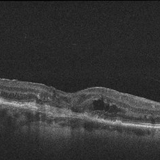
80-year-old female with subfoveal occult CNV and large extrafoveal PED which underwent spontaneous RPE rip. OCT shows subfoveal CNV and intraretinal cystic edema
Condition/keywords: pigment epithelial detachment (PED), retinal pigment epithelium (RPE) tear
-
---thumb.jpg/image-square;max$300,300.ImageHandler) Toxocariasis Cyst
Toxocariasis Cyst
Feb 13 2013 by From the Collections of Thomas M. Aaberg, MD and Thomas M. Aaberg Jr., MD
Mid-peripheral retinal cyst.
Condition/keywords: retinal cyst, toxocariasis
-
 Chronic Retinal Detachment: Features Slide 3
Oct 22 2012 by Ronald C. Gentile, MD
Chronic retinal detachments can be associated with demarcation lines (tidemarks), subretinal bands or sheets, and retinal cysts. Fundus photo of a chronic retinal detachment reveals a retinal cyst within the peripherally detached temporal retina.
Condition/keywords: chronic retinal detachment
-
---thumb.jpg/image-square;max$300,300.ImageHandler) Toxocariasis Cyst
Toxocariasis Cyst
Feb 13 2013 by From the Collections of Thomas M. Aaberg, MD and Thomas M. Aaberg Jr., MD
Mid-peripheral retinal cyst.
Condition/keywords: retinal cyst, toxocariasis
-
 Retinal Detachment
Nov 9 2012 by Norman Byer
This 18-year-old girl gave the history of having been hit in this eye three years before with a fist and of having retinal surgery nine months previously, which was temporarily successful. When the photograph was taken, she had a total left retinal detachment with a small nasal dialysis which had not been treated. She also had two prominent intraretinal cysts, one of which is shown here. The retina promptly reattached following further surgery and the next slide shows an interesting change in this cyst.
Condition/keywords: intraretinal cyst, small nasal dialysis
-
 Retinal Detachment
Nov 9 2012 by Norman Byer
This is the same lesion as in the previous photograph shown 13 days after surgery. Not only is the retina reattached, but the cyst has now completely disappeared even though no treatment of any kind was applied in the vicinity of the cyst. Long detached retinas tend to develop intraretinal cysts, and these tend to disappear following reattachment of the retina even without direct treatment to the cysts.
Condition/keywords: intraretinal cyst, re-attached retinal detachment (RRD)
-
---thumb.jpg/image-square;max$300,300.ImageHandler) Retinal Cyst?
Retinal Cyst?
Apr 4 2014 by H. Michael Lambert, MD
cystic structure in under retina. Etiology?
Condition/keywords: retinal cyst
-
 Cystercercosis
Sep 17 2012 by Prema Abraham, MD
Fundus photograph of 25-year-old male with subretinal cystercercosis.
Photographer: Dan Parks, Black Hills Regional Eye Institute, Rapid City South Dakota
Imaging device: Topcon 50 EX
Condition/keywords: cysticercosis
-
 Punctate Inner Choroidopathy Complicated with CNV
Jun 5 2013 by Henry J. Kaplan, MD
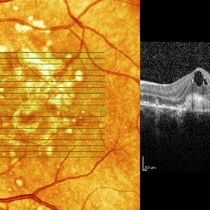
OCT of the same patient demonstrates CNV complex with intraretinal cystoid edema #3.
Photographer: Angela Andersson
Imaging device: HRA II
Condition/keywords: choroidal neovascularization (CNV), punctate inner choroidopathy (PIC)
-
 Multiple Retinal Cysts Associated With Chronic Retinal Detachment
Sep 24 2018 by samarth mishra
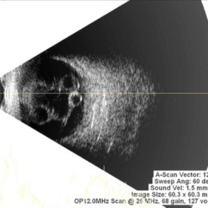
Patient presented with a diminution of vision in left eye since few months. On B-scan ultrasonography multiple retinal cysts with a total retinal detachment were noted.
Photographer: Aditya Birla Sankara Nethralaya, West Bengal , Kolkata , India
Condition/keywords: B scan ultrasound, chronic retinal detachment, intraretinal cyst, retinal cyst
-
 Multiple Retinal Cysts Associated With Chronic Retinal Detachment
Sep 24 2018 by samarth mishra
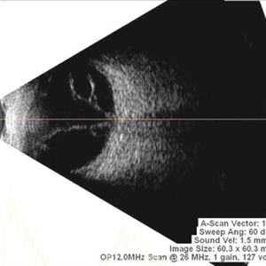
Patient presented with a diminution of vision in left eye since few months. On B-scan ultrasonography multiple retinal cysts with a total retinal detachment were noted.
Photographer: Aditya Birla Sankara Nethralaya, West Bengal , Kolkata , India
Condition/keywords: B scan ultrasound, chronic retinal detachment, intraretinal cyst, retinal cyst
-
 Multiple Retinal Cysts Associated With Chronic Retinal Detachment
Sep 24 2018 by samarth mishra
Patient presented with a diminution of vision in left eye since few months. On B-scan ultrasonography multiple retinal cysts with a total retinal detachment were noted.
Photographer: Aditya Birla Sankara Nethralaya, West Bengal , Kolkata , India
Condition/keywords: B scan ultrasound, chronic retinal detachment, intraretinal cyst, retinal cyst
-
 Cystercercosis
Sep 17 2012 by Prema Abraham, MD
Fundus photgraph of 25-year-old male with subretinal cystercercosis.
Photographer: Dan Parks, Black Hills Regional Eye Institute, Rapid City South Dakota
Imaging device: Topcon 50EX
Condition/keywords: cysticercosis
-
 Cystercercosis
Sep 17 2012 by Prema Abraham, MD
Fundus photograph of 25-year-old male with subretinal cystercercosis.
Photographer: Dan Parks, Black Hills Regional Eye Institute, Rapid City South Dakota
Imaging device: Topcon 50EX
Condition/keywords: cysticercosis
-
 Rhegmatogenous Retinal Detachment With Retinal Dialysis and Intraretinal Cyst
Mar 18 2020 by Giridhar Anantharaman, MS
Optos ultra-widefield retinal imaging of the left eye of a 30-year-old lady with rhegmatogenous retinal detachment with inferotemporal retinal dialysis and a large intraretinal cyst.
Photographer: Rakesh PR, Giridhar Eye Institute, Kerala, India
Imaging device: Optos UWF Daytona plus
Condition/keywords: intraretinal cyst, retinal dialysis
-
 Optic Nerve Pit / Serous Detachment
Feb 20 2013 by From the Collections of Thomas M. Aaberg, MD and Thomas M. Aaberg Jr., MD
No history; FA; serous detachment intraretinal cyst; lamellar hole.
Condition/keywords: intraretinal cyst, optic nerve pit
-
 Optic Nerve Pit / Serous Detachment
Feb 20 2013 by From the Collections of Thomas M. Aaberg, MD and Thomas M. Aaberg Jr., MD
No history; FA; serous detachment intraretinal cyst; lamellar hole.
Condition/keywords: intraretinal cyst, optic nerve pit
-
 Multiple Retinal Cysts Associated With Chronic Retinal Detachment
Sep 24 2018 by samarth mishra
Patient presented with a diminution of vision in left eye since few months. On B-scan ultrasonography multiple retinal cysts with a total retinal detachment were noted.
Photographer: Aditya Birla Sankara Nethralaya, West Bengal , Kolkata , India
Condition/keywords: B scan ultrasound, chronic retinal detachment, intraretinal cyst, retinal cyst
-
 Documented Retinal Pars Plana Cysts
Mar 14 2018 by Asaf Friehmann
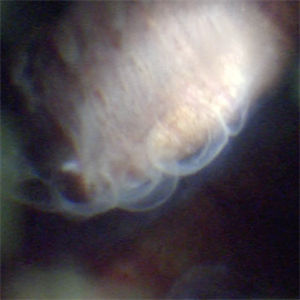
Photograph taken during indentation of a 74-year-old patient who underwent a 25G pars plana vitrectomy (PPV) for repair of dislocated IOL, when this rarely documented peripheral retinal cyst which was found.
Photographer: Alexander Rubowitz
Condition/keywords: peripheral retinal cyst
-
 Subretinal Cysticercosis
Sep 10 2020 by Anamika Dwivedi
Fundus photograph of a 22-year-old male, who was referred from physician as a case of headache under evaluation, with a history of headache for 1 month. Fundus showing bilateral subretinal cysticercus cyst with scolex.
Photographer: Dr Anamika Dwivedi
Imaging device: topcon
Condition/keywords: bilateral subretinal cysticercosis
 Loading…