Search results (61 results)
-
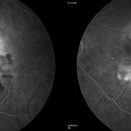
 Fibrovascular PED
Feb 21 2014 by Roy Schwartz, MD
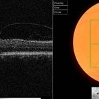
72-year-old female with fibrovascular PED. Upper picture - PED with sub RPE hyper-reflective substance, in a multi-layered pattern, corresponding to fibrovascular PED. CME. Lower picture - PED flattened, a denser sub RPE hyperreflective substance is seen. CME resolved.
Condition/keywords: fibrovascular pigment epithelial detachment (PED), neovascular age-related macular degeneration (AMD), optical coherence tomography (OCT), ranibizumab
-
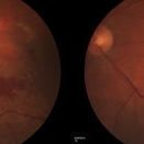
 Active Neovascular AMD With Disciform Scar
Apr 30 2015 by Mitzy E Torres Soriano, MD
Active neovascular AMD with disciform scar.
Photographer: Mitzy E. Torres Soriano, MD; Centro medico Cagua-Estado Aragua. Venezuela
Imaging device: TOPCON
Condition/keywords: disciform scar, disciform with hemorrhage, neovascular age-related macular degeneration (AMD), wet age-related macular degeneration (wet AMD)
-
 Vitreomacular Traction and AMD
Sep 15 2012 by Hamid Ahmadieh, MD
Fundus photograph and OCT image of a 70-year-old man with a subfoveal CNV scar secondary to neovascular AMD and a vitreomacular adhesion.
Photographer: Hamid Ahmadieh, MD, Ophthalmic Research Center, Labbafinejad Medical Center, Shahid Beheshti University of Medical Sciences
Imaging device: Topcon OCT
Condition/keywords: choroidal neovascularization (CNV), neovascular age-related macular degeneration (AMD), vitreomacular adhesion, vitreomacular traction (VMT)
-
 Submacular Hemorrhage
Apr 24 2018 by Pauline T Merrill, MD, FASRS
Fundus photo of left eye of a 65-year-old AMD patient who presented with sudden drop of vision from 20/30 to CF due to a large submacular hemorrhage, 7 months following her last Eylea injection. She underwent immediate injection of C3F8 in the office, with little effect. 10 days later vitrectomy with subretinal tPA and air-fluid exchange was performed, with successful displacement of the hemorrhage.
Photographer: Ermelinda Diaz, Illinois Retina Associates, Chicago, Illinois
Imaging device: Topcon 50DX
Condition/keywords: neovascular age-related macular degeneration (AMD), submacular hemorrhage
-
 Neovascular AMD with Active CNV
Jan 2 2018 by Carolyn Daley
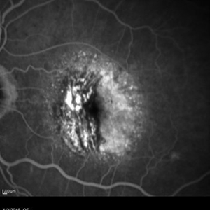
30 degree fluorescein angiogram of an 80-year-old woman with neovascular AMD with active CNV in the left eye. Patient is being treated with Avastin.
Photographer: Carolyn Daley, Retina Specialists of Michigan
Imaging device: Heidelberg Spectralis
Condition/keywords: 30 degrees, choroidal neovascularization (CNV), Heidelburg Spectralis, left eye, neovascular age-related macular degeneration (AMD)
-
 Neovascular ARMD With Subretinal Hemorrhage, Red-Free Photos - Stereo
Nov 26 2014 by James B. Soque, CRA, OCT-C, COA, FOPS
Stereo FC, RF and FA of a 77-year-old white female with visual acuity CC 20/200-3, with left eye neovascular ARMD, drusen, and subretinal hemorrhage with hard exudates temporally. Peripheral retina reveals cobblestone degeneration.
Photographer: James Soque, CRA, COA, Island Retina, Shirley, NY
Imaging device: Topcon TRC 50 EX, with MERGE software and OIS 5 MP digital Camera
Condition/keywords: neovascular age-related macular degeneration (AMD), red-free, stereo pair
-
 Subretinal Hemorrhage Due to SRNVM, Fluorescein Angiogram Photograph
Dec 1 2016 by James B. Soque, CRA, OCT-C, COA, FOPS
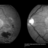
89-year-old white male with NVAMD and new subretinal hemorrhage, fluorescein angiogram, early phase, of the right eye. Currently receiving anti VEGF treatment.
Photographer: James Soque, CRA, OCT-C, COA, Island Retina, Shirley, NY
Imaging device: Topcon TRC 50 DX, with MERGE software
Condition/keywords: hemorrhage, Hot spot, neovascular age-related macular degeneration (AMD), subretinal hemorrhage, subretinal blood, wet age-related macular degeneration (wet AMD)
-
 Neovascular ARMD With Subretinal Hemorrhage, Fluorescein Angiography Photos - Stereo
Oct 14 2014 by James B. Soque, CRA, OCT-C, COA, FOPS
Stereo FC, RF and FA of a 77-year-old white female with visual acuity CC 20/200-3, with left eye neovascular ARMD, drusen, and subretinal hemorrhage with hard exudates temporally. Peripheral retina reveals cobblestone degeneration.
Photographer: James Soque, CRA, COA, Island Retina, Shirley, NY
Imaging device: Topcon TRC 50 EX, with MERGE software and OIS 5 MP digital Camera
Condition/keywords: neovascular age-related macular degeneration (AMD), stereo pair
-
 RAP Lesions
Sep 29 2014 by Thomas A. Ciulla, MD, MBA, FASRS
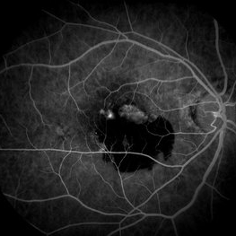
Fluorescein angiogram of an 81-year-old man revealing several RAP lesions superior to fovea.
Photographer: Stuart Alfred CRA
Condition/keywords: choroidal neovascular membrane (CNVM), neovascular age-related macular degeneration (AMD), retinal angiomatous proliferation (RAP), wet age-related macular degeneration (wet AMD)
-
 Chronical Submacular Hemorrhage in the Setting of Neovascular AMD
Mar 23 2015 by Rita Couceiro, MD, MS
An 80-year-old male, with a history of hypertension and high cholesterol, complained of acute and painless vision loss in his left eye (OS) in the previous 5 months. On observation best corrected visual acuity in OS was hand motion. A dense vitreous opacity in OS precluded fundus examination. Ocular ultrasound revealed vitreous hemorrhage and thickening of the macular area. The patient was submitted to pars plana vitrectomy, which disclosed a large submacular hemorrhage with chronical features and disciform scarring in the setting of neovascular AMD.
Imaging device: Intraoperative fundus photograph
Condition/keywords: neovascular age-related macular degeneration (AMD), submacular hemorrhage, wet age-related macular degeneration (wet AMD)
-
 Neovascular AMD
Jan 3 2017 by Jason Griffith
65-year-old female dx neovascular AMD with active CNV.
Photographer: Jason Griffith, Tennessee Retina
Imaging device: Topcon TRC-50EX
Condition/keywords: choroidal neovascularization (CNV), neovascular age-related macular degeneration (AMD)
-
 Neovascular AMD - Classic CNV FA
Aug 7 2013 by H. Michael Lambert, MD
Neovascular AMD - classic CNV FA
Condition/keywords: choroidal neovascularization (CNV), neovascular age-related macular degeneration (AMD)
-
 Neovascular AMD
Mar 17 2015 by Jason Griffith
Fundus photograph of a 72-year-old female with neovascular AMD.
Photographer: Jason Griffith, Tennessee Retina, Nashville, TN
Imaging device: Topcon TRC-50EX
Condition/keywords: neovascular age-related macular degeneration (AMD)
-
 ARMD
ARMD
Aug 7 2013 by H. Michael Lambert, MD
Subretinal hemorrhage,
Condition/keywords: neovascular age-related macular degeneration (AMD), subretinal hemorrhage
-
 Neovascular AMD
Mar 17 2015 by Jason Griffith
Photograph of a 72-year-old female with neovascular AMD.
Photographer: Jason Griffith, Tennessee Retina, Nashville, TN
Condition/keywords: neovascular age-related macular degeneration (AMD)
-
 Neovascular AMD
Mar 17 2015 by Jason Griffith
Photograph of a 72-year-old female with neovascular AMD.
Photographer: Jason Griffith, Tennessee Retina, Nashville, TN
Condition/keywords: neovascular age-related macular degeneration (AMD)
-
 ARMD
ARMD
Aug 7 2013 by H. Michael Lambert, MD
ARMD, laser to CNV, 3/22/90.
Photographer: Donald Lowd
Condition/keywords: neovascular age-related macular degeneration (AMD)
-
 ARMD
ARMD
Aug 7 2013 by H. Michael Lambert, MD
ARMD, one month post laser to perifoveal CNV 4/90
Condition/keywords: neovascular age-related macular degeneration (AMD)
-
 Submacular Hemorrhage
Apr 24 2018 by Pauline T Merrill, MD, FASRS
Fundus photo of left eye of a 65-year-old AMD patient, 3 days following injection of C3F8 and face-down positioning; there was little change in the large submacular hemorrhage. There was no further change one week later, at which time the patient elected surgery.
Photographer: Karen Parque, Illinois Retina Associates, Chicago, Illinois
Imaging device: Topcon 50DX
Condition/keywords: neovascular age-related macular degeneration (AMD), submacular hemorrhage
-
 ARMD, SRNVM
Aug 7 2013 by H. Michael Lambert, MD
ARMD, SRNVM with sub retinal hemorrhage, 79-year-old white female, 20/300.
Condition/keywords: neovascular age-related macular degeneration (AMD), subretinal hemorrhage
-
 Neovascular AMD
Jan 3 2017 by Jason Griffith
75-year-old male with neovascular AMD with disciform scar OS
Photographer: Jason Griffith
Imaging device: Topcon TRC-50EX
Condition/keywords: disciform scar, neovascular age-related macular degeneration (AMD)
-
 Neovascular ARMD With Subretinal Hemorrhage, Fundus Color Photos- Stereo
Oct 14 2014 by James B. Soque, CRA, OCT-C, COA, FOPS
Stereo FC, RF and FA of a 77-year-old white female with visual acuity CC 20/200-3, with left eye neovascular ARMD, drusen, and subretinal hemorrhage with hard exudates temporally. Peripheral retina reveals cobblestone degeneration.
Photographer: James Soque, CRA, COA, Island Retina, Shirley, NY
Imaging device: Topcon TRC 50 EX, with MERGE software and OIS 5 MP digital Camera
Condition/keywords: fundus photograph, neovascular age-related macular degeneration (AMD), stereo pair
-
 Silicone Oil Droplet After Avastin Injection
Aug 23 2016 by Roger A. Goldberg, MD, MBA
Silicone oil droplet in the eye of a patient being treated with Avastin for neovascular AMD (August 2016).
Photographer: Alexandra Estrella
Imaging device: Zeiss FF450
Condition/keywords: neovascular age-related macular degeneration (AMD), silicone oil
-
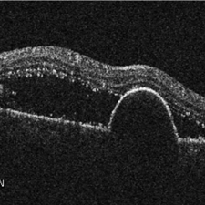 Retinal Pigment Epithelium Detachment
Jan 26 2016 by Andrea Arriola-Lopez, MD MSc
89-year-old woman, VA 20/800, IOP 13 mmHg. OCT showed subretinal fluid and PED. Anti-VEGF was administrated.
Photographer: Andrea Elizabeth Arriola MD, MSc
Imaging device: Cirrus
Condition/keywords: neovascular age-related macular degeneration (AMD), neovascularization (NV), sub-retinal pigment epithelium (RPE)
-
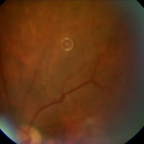
 ARMD with perifoveal CNV
Aug 7 2013 by H. Michael Lambert, MD
ARMD with CNV OD pre-laser 3/22/90
Condition/keywords: neovascular age-related macular degeneration (AMD)
 Loading…