Search results (609 results)
-
 Ozurdex implant
Aug 23 2012 by Daniel A. Adelberg, MD, FASRS
Anterior Segment photograph of a 50 year old with Uveitis and Cystoid Macular Edema status post Intravitreal injection of an Ozurdex dexamethasone implant
Photographer: Robert Ramsey, Southwestern Eye Center, Mesa Arizona
Condition/keywords: Ozurdex implant
-
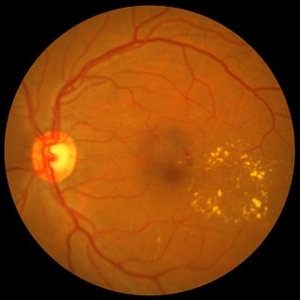 NPDR with CSME
Oct 8 2012 by Jeffrey G. Gross, MD, FASRS
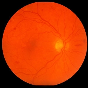
NPDR with CSME with circinate ring of lipid s/p laser.
Condition/keywords: circinate ring, laser, macular edema, nonproliferative diabetic retinopathy
-
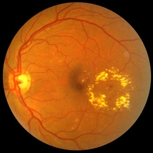 NPDR with CSME
Oct 8 2012 by Jeffrey G. Gross, MD, FASRS
NPDR with CSME with circinate ring of lipid.
Condition/keywords: circinate ring, macular edema, nonproliferative diabetic retinopathy
-
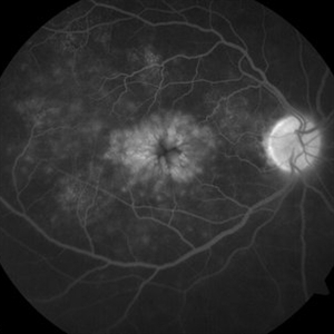
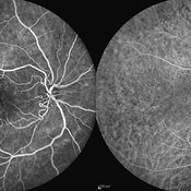
 Diabetic Macular Edema, Proliferative Diabetic Retinopathy, Neovascularization Elsewhere, DME, PDR, NVE
Apr 1 2013 by James B. Soque, CRA, OCT-C, COA, FOPS
39-year-old white female and long standing diabetis, c/o new peripheral symptoms of left eye. FA OS reveals diabetic macular edema, microaneurysms, and neovasculaization elsewhere. Fluorescein Angogram, Early Phase, 50 Deg, 2x Mag.
Photographer: James B Soque, CRA, COA
Imaging device: Topcon TRC 50DX with MERGE software, OIS 10.6.45
Condition/keywords: diabetic macular edema, neovascularization (NV), proliferative diabetic retinopathy (PDR)
-
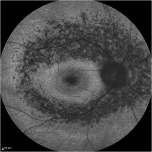 Retinitis Pigmentosa - Fundus Autofluorescence
Sep 20 2014 by Rameez N Hussain, MD
Fundus autofluorescence of retinitis pigmentosa showing hyperautofluorescent rings or foveal hyperautofluorescence.
Photographer: Dr.Rameez N Hussain, MD, Central Imaging Center, Vitreo Retinal Services, Giridhar Eye Institute, Cochin, India
Imaging device: Heidelberg Blue Peak Autofluorescence imaging.
Condition/keywords: bone spicule, cystoid macular edema (CME), fundus autofluorescence (FAF), retinitis pigmentosa
-
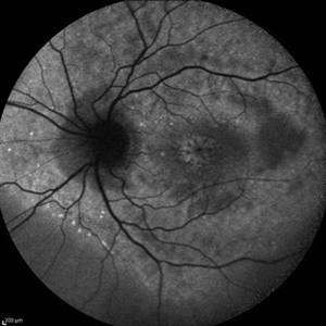 Cystoid Macular Edema (CME)
Sep 11 2012 by Hamid Ahmadieh, MD
Autofluorescence imaging of the left eye of a 17-year-old boy with chronic intermediate uveitis showing CME.
Photographer: Hamid Ahmadieh, MD, Ophthalmic Research Center, Labbafinejad Medical Center, Shahid Beheshti University of Medical Sciences
Imaging device: Heidelberg Spectralis
Condition/keywords: autofluorescence imaging, cystoid macular edema (CME), intermediate uveitis
-
 Branch Retinal Vein Occlusion with Macular Edema
Aug 23 2012 by Gerardo Garcia-Aguirre, MD
Fundus photograph composition of the left eye, showing flame-shaped and blot hemorrhages in the superotemporal quadrant, with hard exudates surrounding the fovea.
Photographer: Noemí Hernández, Asociación para Evitar la Ceguera en México
Condition/keywords: branch retinal vein occlusion (BRVO), macular edema
-
 NPDR with CSME
Oct 8 2012 by Jeffrey G. Gross, MD, FASRS
NPDR with CSME with circinate ring of lipid.
Condition/keywords: circinate ring, macular edema, nonproliferative diabetic retinopathy
-
 Hypertensive Retinopathy
Feb 25 2013 by Suber S. Huang, MD, MBA, FASRS
32-year-old African American male with Grade IV hypertensive retinopathy and acute renal failure. Vision OD 20/70, OS 20/25. Creatine 7.1. BP: 250/150.
Photographer: Geoffrey Pankhurst, University Hospitals, Eye Institute/Dept. Ophthalmology and Visual Sciences Case Western Reserve University Cleveland, OH
Imaging device: Topcon TRC 50x
Condition/keywords: acute renal failure, disc edema, exudate, hypertension, hypertensive retinopathy, ischemia, macular edema, macular ischemia, optic disc edema
-
 BSC CME OS
Nov 10 2012 by Pauline T Merrill, MD, FASRS
Fundus photograph left eye of a 42-year-old Caucasian male with birdshot retinochoroidopathy (HLA-A29+) and cystoid macular edema (CME)
Condition/keywords: birdshot retinochoroidopathy, cystoid macular edema (CME), posterior uveitis, uveitis
-
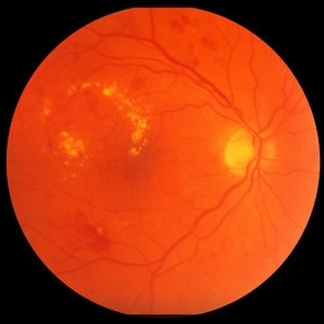 Nonproliferative Diabetic Retinopathy with Clinically Significant Macular Edema and Circinate Lipid Ring
Oct 15 2012 by Jeffrey G. Gross, MD, FASRS
NPDR with CSME and circinate lipid ring.
Condition/keywords: circinate lipid ring, clinically significant macular edema (CSME), macular edema, nonproliferative diabetic retinopathy
-
 Diabetic Retinopathy Hard Exudates OS
Jun 30 2013 by Rogerio N Shinsato, MD, PhD
Fundus photograph with diabetic retinopathy.
Condition/keywords: diabetic macular edema, foveal hard exudates
-
 Hypertensive Retinopathy
Feb 25 2013 by Suber S. Huang, MD, MBA, FASRS
32-year-old African American male with Grade IV hypertensive retinopathy and acute renal failure. Vision OD 20/70, OS 20/25. Creatine 7.1. BP: 250/150.
Photographer: Geoffrey Pankhurst, University Hospitals, Eye Institute/Dept. Ophthalmology and Visual Sciences Case Western Reserve University Cleveland, OH
Imaging device: Topcon TRC 50x
Condition/keywords: acute renal failure, disc edema, exudate, hypertension, hypertensive retinopathy, ischemia, macular edema, macular ischemia, optic disc edema
-
 CME-FFA
Apr 28 2015 by Neha Goel, MS DNB FRCS (Glasg)
Fundus fluorescein angiography of the right eye showing flower-petal appearance of the leakage.
Photographer: Neha Goel
Imaging device: Zeiss visucam
Condition/keywords: cystoid macular edema (CME)
-
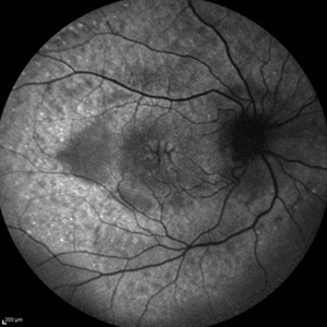 Cystoid Macular Edema (CME)
Sep 11 2012 by Hamid Ahmadieh, MD
Fundus autofluorescence (FAF) of the right eye a 17-year-old boy with chronic intermediate uveitis showing CME.
Photographer: Hamid Ahmadieh, MD, Ophthalmic Research Center, Labbafinejad Medical Center, Shahid Beheshti University of Medical Sciences
Imaging device: Heidelberg Spectralis
Condition/keywords: cystoid macular edema (CME), fundus autofluorescence (FAF), intermediate uveitis
-
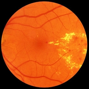
 Diabetic Retinopathy, CSME, Color Fundus Photo
Mar 18 2015 by James B. Soque, CRA, OCT-C, COA, FOPS
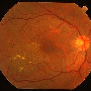
A 58-year-old diabetic male with a longstanding history of diabetic eye disease. Left eye color fundus photo shows extensive CSME, Clinically Significant Macular Edema, with deposits of hard exudates at fixation. There is extensive scattering of hard exudates and sheathing of the vessels.
Photographer: James B Soque, CRA COA
Imaging device: Topcon TRC 50 DX, OIS 5 MP Camera, MERGE software
Condition/keywords: background diabetic retinopathy (BDR), creamy yellow exudates, diabetes, exudates over the posterior pole, neovascularization of the disc (NVD), vessel sheathing
-
 Cistoid Macular Edema (CME) Caused due Branch Retinal Vein Occlusion (BRVO)
Sep 9 2012 by Ratimir Lazic, MD, PhD
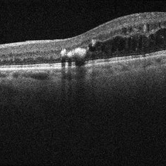
OCT image of a 61 - year - old male with cistoid macular edema due to branch retinal vein occlusion
Photographer: Ratimir Lazic, PhD MD
Imaging device: SOCT Copernicus
Condition/keywords: branch retinal vein occlusion (BRVO)
-
 CRVO-associated Macular Edema
Jul 8 2012 by Jeffrey S. Heier, MD
Young physician with CRVO and macular edema
Imaging device: Spectralis
Condition/keywords: central retinal vein occlusion (CRVO), macular edema
-
 Nonproliferative Diabetic Retinopathy with Clinically Significant Macular Edema and Circinate Lipid Ring
Oct 15 2012 by Jeffrey G. Gross, MD, FASRS
NPDR with CSME and circinate lipid ring.
Condition/keywords: circinate lipid ring, clinically significant macular edema (CSME), macular edema, nonproliferative diabetic retinopathy
-
 Circinate Ring OCT
Aug 24 2012 by John S. King, MD
DR; hard exudate at edges of macular edema and are hyper-reflective with shadowing.
Photographer: Kristin Konecki, OcuSight Eye Care Center, Rochester, NY
Condition/keywords: circinate ring
-
 Cystoid Macular Edema (CME)
Sep 11 2012 by Hamid Ahmadieh, MD
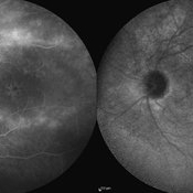
Late phase FA & ICG angiography imagings of the left eye a 17-year-old boy with CME & retinal periphlebitis secondary to chronic intermediate uveitis.
Photographer: Hamid Ahmadieh, MD, Ophthalmic Research Center, Labbafinejad Medical Center, Shahid Beheshti University of Medical Sciences
Imaging device: Heidelberg Spectralis
Condition/keywords: cystoid macular edema (CME), indocyanine green (ICG) angiography, intermediate uveitis
-
 MPC for CSME
Mar 29 2013 by Henry J. Kaplan, MD
Right after MPC for CSME in diabetes (before the introduction of anti-VEGFs).
Condition/keywords: clinically significant macular edema (CSME), diabetic macular edema, multifocal chorioretinitis (MCP)
-
 Double Macular Branch Retinal Vein Occlusion
Sep 22 2014 by Mallika Goyal, MD
Left fundus of a 53-year-old lady with dyslipidemia who presented with left eye sudden vision drop 5 days prior to presenting. There is superior and inferior macular branch retinal vein occlusion. She had macular edema, and was treated with intravitreal Avastin.
Photographer: Mallika Goyal, MD, Apollo Health City, Jubilee Hills, Hyderabad-500033
Condition/keywords: macular branch retinal vein occlusion (BRVO)
-
---thumb.jpg/image-square;max$300,300.ImageHandler) Diabetic Retinopathy Hard Exudates OD
Diabetic Retinopathy Hard Exudates OD
Jun 30 2013 by Rogerio N Shinsato, MD, PhD
Fundus photograph with diabetic retinopathy.
Condition/keywords: diabetic macular edema, foveal hard exudates
-
 Cystoid Macular Edema (CME)
Sep 11 2012 by Hamid Ahmadieh, MD
FA & ICG angiography imagings of a 17-year-old boy with CME secondary to chronic intermediate uveitis.
Photographer: Hamid Ahmadieh, MD, Ophthalmic Research Center, Labbafinejad Medical Center, Shahid Beheshti University of Medical Sciences
Imaging device: Heidelberg Spectralis
Condition/keywords: cystoid macular edema (CME), indocyanine green (ICG) angiography, intermediate uveitis
 Loading…