Search results (3038 results)
-
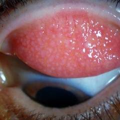
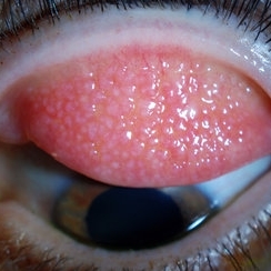 Giant Papillary Conjunctivitis, Left Upper Eyelid
Jul 22 2013 by Jason S. Calhoun
Contact lens wearer in for exam. Has rough feeling underneath both eyelids. Patient thought it was through SCL wear. Patient VA was 20/20. right eye, 20/30, left eye. Underneath the left upper eyelid, you can see papillary inflammation and redness.
Photographer: Jason S. Calhoun, Department of Ophthalmology, Mayo Clinic Jacksonville, Florida
Imaging device: TOPCON D-90 SL NIKON CAMERA
Condition/keywords: giant papillary conjunctivitis
-
---thumb.jpg/image-square;max$300,300.ImageHandler) CMV Retinitis in a Patient with the Diagnosis of AIDS
CMV Retinitis in a Patient with the Diagnosis of AIDS
Feb 27 2013 by Henry J. Kaplan, MD
CMV retinitis, left eye: classic form in AIDS patient. Hemorrhagic retinitis mainly in the superior arcade.
Condition/keywords: AIDS
-
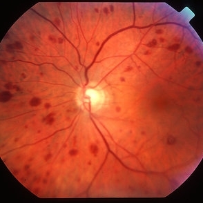 Ocular ischaemic syndrome colour 1
Jan 11 2013 by Alex P. Hunyor, MD
Ocular ischaemic syndrome, left eye - color image, posterior pole. Note: dilated but not tortuous veins, attenuated arteries, and multiple intraretinal haemorrhages.
Condition/keywords: ocular ischemic syndrome
-
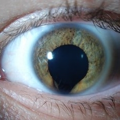 Keyhole Pupil Coloboma
Jul 13 2013 by Jason S. Calhoun
14-year-old male presents with decreased vision in the left eye. Dx with iris and retinal coloboma in the left eye. Patient VA was 20/20, right eye, 20/100 left eye with pinhole improvement 20\50. Patient was fitted for SCL in the left eye.
Photographer: Jason S. Calhoun, Department of Ophthalmology, Mayo Clinic Jacksonville, Florida
Imaging device: TOPCON D-90 SL NIKON CAMERA
Condition/keywords: deformed pupil
-
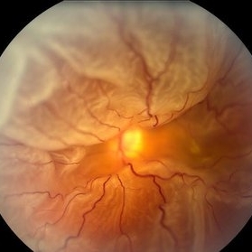
 Toxoplasma Neuroretinitis (Jensen`s Disease)
Feb 25 2013 by Henry J. Kaplan, MD
Toxoplasma neuroretinitis in the left eye of a patient with macular star formation, retinitis adjacent to the optic nerve head with disc swelling.
Condition/keywords: Jensen disease, ocular toxoplasmosis, toxoplasmosis
-
 Total Rhegmatogenous Retinal Detachment With Severe PVR
May 27 2015 by Darin R. Goldman, MD
63-year-old pseudophakic male with hand motion vision in the left eye due to a total retinal detachment with severe proliferative vitreoretinopathy.
Condition/keywords: proliferative vitreoretinopathy (PVR), retinal tear
-
 Venous Loop & Venous Beading
May 31 2014 by Hamid Ahmadieh, MD
Color fundus photograph of the left eye of a diabetic patient with NVD, NVE, venous loop and venous beading.
Photographer: Elham Salehi, Negah Eye Center, Tehran
Condition/keywords: color fundus photograph, neovascularization elsewhere (NVE), neovascularization of the disc (NVD), proliferative diabetic retinopathy (PDR), venous beading, venous loop
-
 Hypertensive Retinopathy Grade IV OS
Mar 13 2013 by Jose Dalma-Weiszhausz, MD
Left eye of young patient with hypertensive retinopathy due to nephrotic syndrome.
Photographer: José Dalma, MD, Dalma & Asoc. Mexico City, Mexico
Condition/keywords: hypertensive retinopathy, renal failure
-
---thumb.JPG/image-square;max$300,300.ImageHandler) Disciform Scar
Disciform Scar
Jul 13 2013 by Jason S. Calhoun
Poor central vision in the left eye due to macular degeneration. Disciform scar.
Photographer: Jason S. Calhoun, Department of Ophthalmology, Mayo Clinic Jacksonville, Florida
Imaging device: TOPCON TRC 50-EX
Condition/keywords: disciform scar, macular degeneration
-
 Ocular Toxocariasis slide 1
Oct 22 2012 by Ronald C. Gentile, MD
40-year-old man from South America was referred for a peripheral retinal scar in his left eye. He had a history of conjunctivitis as a child with exposure to multiple pets (cats and dogs). Fundus photo revealed a peripheral scarred sub-retinal granuloma located superior nasal with a retinal fold and traction extending to the optic nerve.
Photographer: The New York Eye & Ear Infirmary Department of Medical Imaging
Condition/keywords: toxocariasis
-
 Encephalitis with Retinal Cotton Wool Spots
Oct 15 2012 by Jeffrey G. Gross, MD, FASRS
Encephalitis, with retinal cotton wool spots, left eye, 20/30.
Condition/keywords: cotton wool spots, encephalitis, left eye
-
---thumb.jpg/image-square;max$300,300.ImageHandler) Multifocal Choroiditis and Panuveitis Syndrome
Multifocal Choroiditis and Panuveitis Syndrome
Feb 26 2013 by Henry J. Kaplan, MD
Multifocal choroiditis, left eye: multiple punched out scar formations in the posterior pole.
Condition/keywords: multifocal choroiditis, panuveitis
-
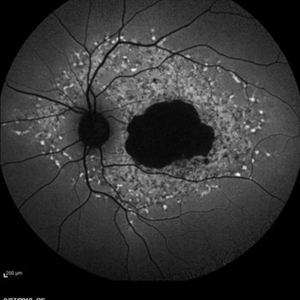
 Macula off Rhegmatogenous Retinal Detachment
Aug 28 2012 by Sharon Fekrat, MD FACS FASRS
62 year old man with a rhegmatogenous retinal detachment involving the foveal center in his left eye as depicted on this Zeiss Stratus OCT image.
Photographer: Michael P. Kelly, FOPS Director, Duke Eye Labs, Duke University Eye Center, Durham, NC
Imaging device: Zeiss Stratus
-
 Giant Papillary Conjunctivitis, Left Upper Eyelid
Jul 22 2013 by Jason S. Calhoun
Contact lens wearer, in for exam. Has rough feeling underneath both eyelids. Patient thought it was through SCL wear. Patient VA was 20/20. right eye, 20/30, left eye. Underneath the left upper eyelid, you can see papillary inflammation and redness.
Photographer: Jason S. Calhoun, Department of Ophthalmology, Mayo Clinic Jacksonville, Florida
Imaging device: TOPCON D-90 SL NIKON CAMERA
Condition/keywords: giant papillary conjunctivitis
-
---thumb.JPG/image-square;max$300,300.ImageHandler) Retinal Coloboma
Retinal Coloboma
Jul 8 2013 by Jason S. Calhoun
14-year-old male with decreased vision in the left eye. Dx with iris and retinal coloboma in the left eye. Patient VA was 20/20, right eye, 20/100 left eye with pinhole improvement 20\50. Patient was fitted for SCL in the left eye.
Photographer: Jason S. Calhoun, Department of Ophthalmology, Mayo Clinic Jacksonville, Florida
Condition/keywords: chorioretinal coloboma
-
 360 Degree Retinal Detachment
Jun 29 2013 by Jason S. Calhoun
Total retinal detachment in the left eye.
Photographer: Jason S. Calhoun, Mayo Clinic Jacksonville, Florida
Imaging device: TOPCON TRC 50-EX
-
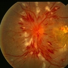
 Hypertensive Retinopathy
Aug 24 2012 by Geoffrey G. Emerson, MD, PhD, FASRS
A 35-year-old man has headaches and decreased vision. The right eye measures 20/25 and the left eye measures 3/200. The blood pressure measures 180/110.
Photographer: Geoffrey Emerson, MD, PhD, Retina Center, Minneapolis
Condition/keywords: hypertensive retinopathy, papilledema, serous retinal detachment
-
 Diabetic Macular Edema, Proliferative Diabetic Retinopathy, Neovascularization Elsewhere, DME, PDR, NVE
Apr 1 2013 by James B. Soque, CRA, OCT-C, COA, FOPS
39-year-old white female and long standing diabetis, c/o new peripheral symptoms of left eye. FA OS reveals diabetic macular edema, microaneurysms, and neovasculaization elsewhere. Fluorescein Angogram, Early Phase, 50 Deg, 2x Mag.
Photographer: James B Soque, CRA, COA
Imaging device: Topcon TRC 50DX with MERGE software, OIS 10.6.45
Condition/keywords: diabetic macular edema, neovascularization (NV), proliferative diabetic retinopathy (PDR)
-
 Myopic CNV
Jan 11 2013 by Alex P. Hunyor, MD
Myopic macular degeneration complicated by subretinal neovascularisation, left eye.
Condition/keywords: high myopia, myopia, myopic choroidal neovascularization (CNV)
-
 Pigmented Peripheral Retinal Degeneration
Jun 27 2013 by Jason S. Calhoun
42-year-old male came in for routine eye exam and to follow up on peripheral retinal degeneration in both eyes. VA is 20/20, right eye and 20/25, left eye. Patient is asymptomatic with no visual complaints.
Photographer: Jason S. Calhoun, Mayo Clinic Jacksonville, Florida
Imaging device: TOPCON TRC 50-EX
Condition/keywords: peripheral retinal degeneration
-
 Late Stage Stargardt's Disease
Mar 13 2013 by Hamid Ahmadieh, MD
Autofluorescence imaging of the left eye of a 46-year-old man with decreased VA due to advanced Stargardt's disease.
Photographer: Nayereh Hadipoor, Negah Eye Center, Tehran
Imaging device: Heidelberg Spectralis
Condition/keywords: autofluorescence imaging, Stargardt disease
-
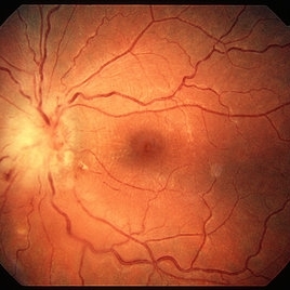
 Epiretinal Membrane
Oct 26 2012 by Sharon Fekrat, MD FACS FASRS
39-year-old female with long standing epiretinal membrane in the left eye and good vision
Photographer: Jim Crowell, Ophthalmic Photographer, Duke Eye Imaging, Durham, NC
Condition/keywords: epiretinal membrane (ERM), macular pucker
-
 White Without Pressure
Jan 31 2018 by Olivia Rainey
Ultra-wide field pseudocolor photograph of a 57-year-old female with white without pressure affecting her left eye. Patient will be having bloodwork done to rule out possible sarcoidosis or sickle cell.
Photographer: Olivia Rainey
Imaging device: Optos
Condition/keywords: blot hemorrhages, color fundus photograph, left eye, Optos, ultra-wide field imaging, white without pressure
-
 Myopic macular degeneration
Jan 11 2013 by Alex P. Hunyor, MD
Myopic macular degeneration, left eye - extensive chorioretinal atrophy.
Condition/keywords: myopic degeneration, myopic fundus, myopic macular degeneration
-
 HIV retinopathy with resolving CMV retinitis - left eye
Jan 11 2013 by Alex P. Hunyor, MD
HIV retinopathy and resolving CMV retinitis, left eye. 36-year-old male with HIV/AIDS. Multiple cotton wool spots due to HIV microangopathy, and an area of resolving CMV retinitis superior to the fovea (patient undergoing treatment with IV ganciclovir).
Condition/keywords: CMV retinitis, HIV retinopathy
 Loading…