Search results (197 results)
-
 Choroidal Melanoma
Jul 4 2012 by John T. Thompson, MD
Amelanotic choroidal melanoma with serous retinal detachment
Condition/keywords: choroidal tumor, exudative retinal detachment, melanoma
-
---thumb.jpg/image-square;max$300,300.ImageHandler) Sturge-Weber Diffuse Hemangioma and Retinal Detachment on B-scan
Sturge-Weber Diffuse Hemangioma and Retinal Detachment on B-scan
Apr 18 2014 by Susanna S. Park, MD, PhD
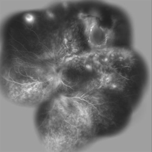
B-scan ultrasonogram of the right eye of an 8 year old Hispanic boy with Sturge -Weber Syndrome showing diffuse choroidal thickening from diffuse choroidal hemangioma and associated total exudative retinal detachment.
Photographer: Ellen Redenbo, University of California Davis Eye Center
Condition/keywords: B scan ultrasound, diffuse choroidal hemangioma, Sturge-Weber syndrome
-
---thumb.jpg/image-square;max$300,300.ImageHandler) Choroidal Metastasis B-Scan
Choroidal Metastasis B-Scan
Jan 10 2014 by Susanna S. Park, MD, PhD
B-scan ultrasound image showing choroidal thickening and exudative retinal detachment in a patient with diffuse choroidal metastasis from breast carcinoma.
Photographer: Ellen Redenbo, University of California Davis Eye Center
Condition/keywords: B scan ultrasound, choroidal metastasis
-
---thumb.jpg/image-square;max$300,300.ImageHandler) vitreous snowballs, peripheral retinal neovascularization, inferior snowbanking, vascular sheathing, and peripheral exudative retinal detachment
vitreous snowballs, peripheral retinal neovascularization, inferior snowbanking, vascular sheathing, and peripheral exudative retinal detachment
Feb 14 2013 by From the Collections of Thomas M. Aaberg, MD and Thomas M. Aaberg Jr., MD
schematic drawing of vitreous snowballs, peripheral retinal neovascularization, inferior snowbanking, vascular sheathing, and peripheral exudative retinal detachment
Condition/keywords: exudative retinal detachment, pars planitis, peripheral retinal neovascularization, snowbank
-
 Disseminated Retinitis and Retinochoroiditis, Metastatic
May 16 2017 by Karen Panzegrau
Fundus photograph of 44-year-old male with plasmacytoma infiltation of the choroid confirmed by biopsy, associated with disseminated retinitis, and retinochoroiditis. Vision is LP. Patient treated with intravitreal methotrexate
Photographer: Karen Panzegrau
Imaging device: Optos
Condition/keywords: metastatic lesion, methotrexate, Optos, plasmacytoma, retinitis, retinochoroiditis, unilateral exudative retinal detachment
-
 Choroidal Melanoma
Jan 30 2019 by Karen Panzegrau
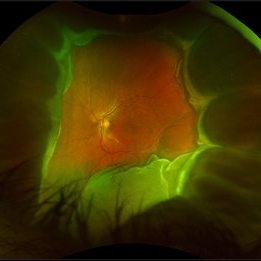
Ultra-wide field optos image of a 27-year-old male patient who presented with loss of vision for about 6-8 weeks. Previous choroidal nevus seen. Recommended annual monitoring. No exam for since 10/2014. Brachytherapy vs enucleation was discussed. Brachytherapy was decided as treatment. Full metastatic work up is being performed.
Photographer: Karen Panzegrau
Imaging device: Optos
Condition/keywords: choroidal nevus, exudative retinal detachment, malignant neoplasm of eye, Optos, ultra-wide field imaging
-
 Von Hippel-Lindau 1
Oct 13 2012 by Hamid Ahmadieh, MD
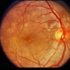
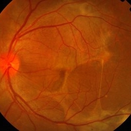
Color fundus photograph of the left eye of a 25-year-old woman with exudative retinal detachment secondary to retinal angiomatosis (Von Hippel-Lindau).
Photographer: Hamid Ahmadieh, MD, Ophthalmic Research Center, Labbafinejad Medical Center, Shahid Beheshti University of Medical Sciences
Imaging device: Topcon Fundus Camera
Condition/keywords: exudative retinal detachment, retinal angiomatous proliferation (RAP), Von Hippel-Lindau
-
 Von Hippel-Lindau
Oct 13 2012 by Hamid Ahmadieh, MD
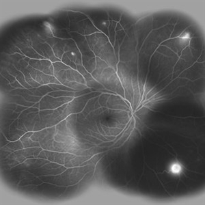
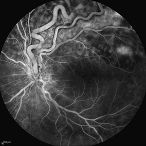
Wide field FA image of the right eye of a 25-year-old woman with retinal angiomatosis (Von Hippel-Lindau). Fundus of the right eye seemed to be normal in ophthalmoscopy.
Photographer: Soodabeh Fooladin, Negah Eye Center, Tehran
Imaging device: Heidelberg Spectralis
Condition/keywords: exudative retinal detachment, retinal angiomatous proliferation (RAP), Von Hippel-Lindau
-
 Amelanotic Choroidal Melanoma
Apr 12 2019 by David L Kilpatrick, MD
Fundus photograph of a 69-year-old male with an amelanotic choroidal melanoma and corresponding exudative retinal detachment. Transvitreal biopsy was performed at the time of radioactive I-125 plaque placement. The genetic expression profile revealed a Class 1A, PRAME negative tumor.
Photographer: Retina Consultants of Alabama, P. C.
Imaging device: Optos
Condition/keywords: amelanotic melanoma
-
 Vogt-Koyanagi-Harada Disease
Feb 20 2015 by H. Michael Lambert, MD
Color photo showing multifocal detachments of the neurosensory retina with underlying cream colored lesions ( possibly Dalen-Fuchs nodules). Large pocket of subretinal fluid in the macula.
Condition/keywords: exudative retinal detachment, Vogt-Koyanagi-Harada
-
---thumb.jpg/image-square;max$300,300.ImageHandler) Harada's with Exudative RD
Harada's with Exudative RD
Oct 13 2012 by Edwin H. Ryan, MD
OCT of a 35-year-old woman with acute vision loss in one eye.
Condition/keywords: exudative retinal detachment, Harada's disease
-
---thumb.JPG/image-square;max$300,300.ImageHandler) Coats Disease
Coats Disease
Oct 11 2012 by Anat Loewenstein, MD
Fluorescein angiography of 6 -year-old girl whose parents have noticed leukocoria in her right eye. On examination severe exudative retinal detachment was diagnosed. On FA of the right eye peripehral capillary non perfusion and peripheral capillary dilatations were seen.
Photographer: Galit Yair-Pur
-
 Coats
Coats
Nov 6 2012 by F. Ryan Prall, MD
22-year-old male with exudative retinal detachment and light-bulb aneurysms seen on FA.
Photographer: Tom Egnatz, Indiana University
Condition/keywords: exudative retinal detachment, light-bulb aneurysms
-
 Von Hippel-Lindau
Oct 13 2012 by Hamid Ahmadieh, MD
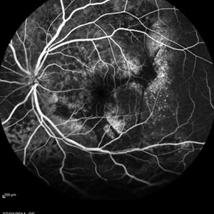
Wide field FA image of the left eye of a 25-year-old woman with exudative retinal detachment secondary to retinal angiomatosis (Von Hippel-Lindau).
Photographer: Soodabeh Fooladin, Negah Eye Center, Tehran
Imaging device: Heidelberg Spectralis
Condition/keywords: exudative retinal detachment, retinal angiomatous proliferation (RAP), Von Hippel-Lindau
-
 Choroidal metastasis case 3 image 1
Jan 11 2013 by Alex P. Hunyor, MD
Large choroidal metastasis from breast carcinoma, with exudative retinal detachment.
Condition/keywords: choroidal metastasis
-
 Harada's with Exudative RD
Oct 13 2012 by Edwin H. Ryan, MD
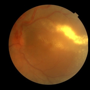
Fundus photograph of a 35-year-old woman with acute vision loss in one eye.
Condition/keywords: exudative retinal detachment, Harada's disease
-
---thumb.jpg/image-square;max$300,300.ImageHandler) Choroidal Metastasis OCT EDI
Choroidal Metastasis OCT EDI
Jan 10 2014 by Susanna S. Park, MD, PhD
Enhanced depth OCT image of the macula of a patient with diffuse choroidal metastasis and exudative retinal detachment from breast carcinoma.
Photographer: Ellen Redenbo, University of California Davis Eye Center
Imaging device: Heidelberg Spectralis
Condition/keywords: choroidal metastasis, enhanced depth imaging, optical coherence tomography (OCT)
-
 Von Hippel-Lindau
Oct 13 2012 by Hamid Ahmadieh, MD
Late FA image of the left eye of a 25-year-old woman with exudative retinal detachment secondary to retinal angiomatosis (Von Hippel-Lindau).
Photographer: Soodabeh Fooladin, Negah Eye Center, Tehran
Imaging device: Heidelberg Spectralis
Condition/keywords: exudative retinal detachment, retinal angiomatous proliferation (RAP)
-
---thumb.jpg/image-square;max$300,300.ImageHandler) Harada's with Exudative RD
Harada's with Exudative RD
Oct 13 2012 by Edwin H. Ryan, MD
OCT of a 35-year-old woman with acute vision loss in one eye.
Condition/keywords: exudative retinal detachment, Harada's disease
-
---thumb.jpg/image-square;max$300,300.ImageHandler) vitreous haze and retinal detachment
vitreous haze and retinal detachment
Feb 14 2013 by From the Collections of Thomas M. Aaberg, MD and Thomas M. Aaberg Jr., MD
color fundus photograph showing vitreous haze and retinal detachment associated with ocular toxoplasmosis.
Condition/keywords: exudative retinal detachment, ocular toxoplasmosis
-
 Multifocal Exudative Detachments Due to VKH
May 14 2014 by Avris Romario Diparaja Siahaan
Fundus Photograph a 38-year-old man with multifocal CSR and inferior exudative retinal detachment on both eyes (Harada Syndrome).
Photographer: Avris Romario Diparaja Siahaan, Klinik Mata Nusantara
Imaging device: Topcon TRC 50 DX Type IA
Condition/keywords: fundus photograph, multifocal central serous chorioretinopathy (CSCR)
-
---thumb.jpg/image-square;max$300,300.ImageHandler) vitreous haze and retinal detachment associated with ocular toxoplasmosis
vitreous haze and retinal detachment associated with ocular toxoplasmosis
Feb 14 2013 by From the Collections of Thomas M. Aaberg, MD and Thomas M. Aaberg Jr., MD
color fundus photograph showing vitreous haze and retinal detachment associated with ocular toxoplasmosis
Condition/keywords: exudative retinal detachment, ocular toxoplasmosis
-
 Uveitis With Exudative Retinal Detachment
May 3 2014 by Mallika Goyal, MD
Fluorescein angiogram of an elderly patient with bilateral posterior uveitis shows punctate hyperfluorescence and inferior RD. He responded well to oral steroids with complete resolution of the uveitis and RD.
Photographer: Mallika Goyal, MD, Apollo Health City, Jubilee Hills, Hyderabad, India
Condition/keywords: exudative retinal detachment, uveitis
-
 Vasoproliferative Tumor With Resultant Total Exudative RD Status Post Vitrectomy/Laser/Oil/Dexamethasone Intravitreal Implant
Apr 25 2017 by Christopher G Fuller, MD
Fundus photograph of a presumptive vasoproliferative tumor (with resultant total exudative retinal detachment) in a 54-year-old white truck driver. Image is taken on post-operative day 4, after 25/27 gauge vitrectomy with drainage retinotomy, air-fluid exchange, endoscopic laser blanching of VPT, oil, and dexamethasone intravitreal implant.
Photographer: Ray Gardner, Texas Retina Associates (Lubbock, TX)
Condition/keywords: exudative retinal detachment
-
 Multifocal Exudative Detachments Due to VKH
May 14 2014 by Avris Romario Diparaja Siahaan
FA (Early Phase) a 38-year-old man with multifocal CSR and inferior exudative retinal detachment on both eyes (Harada Syndrome) with 55 degree lens
Photographer: Avris Romario Diparaja Siahaan, Klinik Mata Nusantara
Imaging device: Heidelberg HRA + OCT Spectralis
Condition/keywords: multifocal central serous chorioretinopathy (CSCR)
 Loading…