File number: 33172
Comments
-
 Gayathri Mohan (November 8 2019)
Have added the details to the description Sir. Also there's an ultrasonography attached to the case.
-
 Suber S. Huang, MD, MBA, FASRS (November 8 2019)
Can you confirm that there is a choroidal mass? These large clear cystic changes can be seen in a variety of conditions. The clinical history lacks critical clinical details. Please add these to the description.
Sign in to comment.
 Initializing download.
-
 By Gayathri Mohan
Co-author(s): Dr. Manish Nagpal, Dr.Navneet Mehrotra- Retina Foundation - Uploaded on Aug 28, 2019.
- Last modified by Gayathri Mohan on Nov 8, 2019.
- Rating
- Appears in
- CHOROIDAL MELANOMA
- Condition/keywords
- choroidal mass, collar button
- Photographer
- Dr. Gayathri Mohan, Retina Foundation
- Imaging device
-
Scanning laser ophthalmoscope
Nidek Mirante SLO - Description
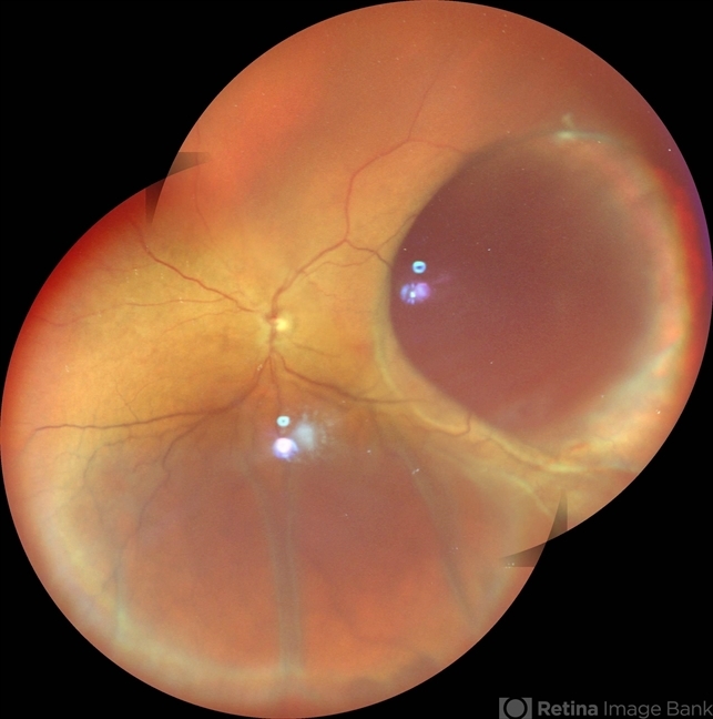
- 50 year old female came with diminution of vision in the LE. Wide field fundus photograph showing an intraocular mass temporally along with an exudative retinal detachment inferiorly. Ultrasonography showed an intraocular mass with collar button appearance suggestive of a Choroidal melanoma. She underwent enucleation and histopathology confirmed a spindle cell choroidal melanoma
---thumb.jpg/image-square;max$79,0.ImageHandler "Vitrectomy Choroidal Mass")


