Search results (44 results)
-
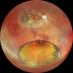
 Optos Picture With Speculum: Dislocated Natural Lens
Oct 9 2018 by John S. King, MD
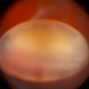
55-year-old white female with history of pathologic myopia+, lattice (laser), SB OU (1990s), and dislocated natural lenses OU that had been watched for years. In the fellow eye she developed phacolytic glaucoma and a PPV, PPL was performed. Plan for both eyes are monitoring. I wanted to get a good picture of her lens today with the optos machine, as the other pics had artifact from the lower lid. It worked out well to use a speculum in the left eye. Vision cc is 20/400 J1+ OD and 20/40 J2 OS; aphakic OU; vitreous clear OD; dislocated lens OS (see pic); retinas attached.
Photographer: Maisee Yang
Imaging device: Optos California
Condition/keywords: dislocated crystalline lens, pathologic myopia, scleral buckle, staphyloma
-
 Aniridia and Dislocated Lens
Oct 18 2012 by Larry Halperin, MD
Aniridia and dislocated lens
Condition/keywords: aniridia, dislocated lens
-
 Sublexed IOL with TID
Dec 13 2013 by Jason S. Calhoun
Patient in with blurred vision in the left eye. Slit lamp exam shows dislocated IOL, inferior, left eye. PXF material on the implant inferiorly.
Photographer: Jason S. Calhoun, Ophthalmic Photographer, Department of Ophthalmology, Mayo Clinic Jacksonville
Imaging device: TOPCON D-90 SL NIKON CAMERA
Condition/keywords: dislocated lens
-
 Lens Luxation
Aug 29 2016 by JEFFERSON R SOUSA, Tecg.º (Biomedical Systems Technology)
Patient, 65-years-old, male, suffered trauma blunt (the jackpot) in the right eye. Ultrasound of the eye found dislocation the total of the crystalline.
Photographer: JEFFERSON R SOUSA - Institute Dr. Suel Abujamra / São Paulo - Brazil
Imaging device: Topcon TRC-50VT, Film Kodak Ektachrome 160 - ASA 100 / 35mm, field of 35 degrees. Flash 100.
Condition/keywords: dislocated lens, lens luxation
-
 Dislocated Lens With Retinal Detachment
Feb 20 2015 by H. Michael Lambert, MD
color photo of Dislocated lens with retinal detachment
Condition/keywords: dislocated lens
-
 Ectopia Lentis
Jan 21 2021 by Jamin S. Brown, MD
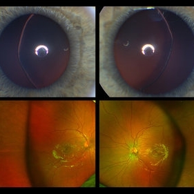
This image serial demonstrates a patient with simple ectopia lentis. Anterior segment photographs in the upper panel demonstrate nasally subluxated crystalline lenses. Widefield fundus photography shows a "pseudo-buckle" which is the result of an optical effect due to the lens subluxation (artifactual image enlargement). Also note the juvenile macular reflex in this young patient. Ectopia lentis can present isolated ("simple") or in combination with various systemic defects (Marfan's syndrome, Weil-Marchesani syndrome or Ehlers-Danlos syndrome to name a few). Isolated ectopia lentis can be hereditary and causative genes have been identified as ADAMTSL4 located on chromosome 4 and FBN1 gene located on chromosome 15. Defects in the genes cause weakness in the zonular fibers which can lead to lens dislocation. Lastly, various ocular disorders such as Aniridia, Axenfeld-Rieger, Pseudoexfoliation or Trauma may also result in lens dislocation or subluxation.
Photographer: Stefanie Palmer CRA, Retina Vitreous Surgeons of CNY
Condition/keywords: dislocated lens, ectopia lentis
-
 Dislocated Lens With Retinal Detachment
Feb 20 2015 by H. Michael Lambert, MD
color photo of Dislocated lens with retinal detachment
Condition/keywords: dislocated lens
-
 Dislocated Lens With Retinal Detachment
Feb 20 2015 by H. Michael Lambert, MD
color photo of Dislocated lens with retinal detachment
Condition/keywords: dislocated lens
-
 Dislocated Lens With Retinal Detachment
Feb 20 2015 by H. Michael Lambert, MD
color photo of Dislocated lens with retinal detachment
Condition/keywords: dislocated lens
-
 Dislocated Lens With Retinal Detachment
Feb 20 2015 by H. Michael Lambert, MD
color photo of Dislocated lens with retinal detachment
Condition/keywords: dislocated lens
-
 Dislocated Lens
Jun 29 2013 by Jason S. Calhoun
84-year-old female comes in with blurred vision in the left eye. VA was 20/30, right eye and count fingers in the left eye. Fundus examination reveals dislocation of the IOL into the vitreous inferiorily at 6-o'clock. Suggest surgery to fix the problem.
Photographer: Jason S. Calhoun, Mayo Clinic Jacksonville, Florida
Imaging device: TOPCON TRC 50-EX
Condition/keywords: dislocated posterior chamber intraocular lens (PCIOL)
-
 Pseudoexfoliation With Partial Subluxation
Jul 12 2013 by Jason S. Calhoun
81-year-old female woke up with loss of vision in the left eye. Patient was hand motion in the left eye. Slit lamp examination shows dislocated lens with pseudoexfoliation material around the iris rim. Proceeded with cataract surgery.
Photographer: Jason S. Calhoun, Department of Ophthalmology, Mayo Clinic Jacksonville, Florida
Condition/keywords: pseudoexfoliation of lens capsule, subluxation of lens
-
---thumb.JPG/image-square;max$300,300.ImageHandler) Dislocated Lens
Dislocated Lens
Jun 30 2013 by Jason S. Calhoun
Dislocation of intra ocular lens into the vitreous, inferiorly.
Photographer: Jason S. Calhoun, Mayo Clinic Jacksonville, Florida
Condition/keywords: dislocated posterior chamber intraocular lens (PCIOL)
-
 Dislocated Lens
Sep 7 2015 by Andrea Arriola-Lopez, MD MSc
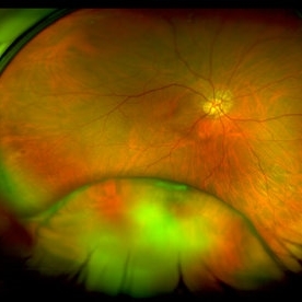
Color fundus photography of right eye of a 54-year-old man, with history of blunt trauma seven month ago. VA HM. IOP 18mmHg. There is no peripherical lesions or traction.
Photographer: Andrea Elizabeth Arriola López, MD, MSc
Imaging device: OPTOS Dakota
Condition/keywords: blunt trauma, dislocated crystalline lens, lens dislocation
-
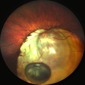
 Bullous RD With Dislocated Lens
Apr 3 2018 by Navneet Mehrotra, DNB
Dislocated clear lens and associated retinal detachment in a young patient with Marfan's syndrome.
Photographer: Navneet Mehrotra
Imaging device: Sony 3 chip camera
Condition/keywords: dislocated crystalline lens, Marfan's syndrome
-
 Pseudoexfoliation With Partial Subluxation
Jul 12 2013 by Jason S. Calhoun
81-year-old female who woke up with loss of vision in the left eye. Patient was hand motion in the left eye. Slit lamp examination shows dislocated lens with pseudoexfoliation material around the iris rim. Proceeded with cataract surgery.
Photographer: Jason S. Calhoun, Department of Ophthalmology, Mayo Clinic Jacksonville, Florida
Condition/keywords: pseudoexfoliation of lens capsule, subluxation of lens
-
---thumb.JPG/image-square;max$300,300.ImageHandler) Dislocated Lens
Dislocated Lens
Jul 14 2013 by Jason S. Calhoun
Patient fell and IOL dislocated to anterior chamber. IOL was placed back after dilation.
Photographer: Jason S. Calhoun, Department of Ophthalmology, Mayo Clinic Jacksonville, Florida
Imaging device: TOPCON D-90 SL NIKON CAMERA
Condition/keywords: anterior dislocation of lens
-
 Slide 7-77
Feb 25 2019 by Lancaster Course in Ophthalmology
Lens dislocated into the anterior chamber.
Condition/keywords: dislocated lens, lens
-
 Pseudoexfoliation With Partial Subluxation
Jul 12 2013 by Jason S. Calhoun
81-year-old female who woke up with loss of vision in the left eye. Patient was hand motion in the left eye. Slit lamp examination shows dislocated lens with pseudoexfoliation material around the iris rim. Proceeded with cataract surgery.
Photographer: Jason S. Calhoun, Department of Ophthalmology, Mayo Clinic Jacksonville, Florida
Condition/keywords: pseudoexfoliation of lens capsule, subluxation of lens
-
 Ultra-Widefield Montage of Traumatically Dislocated Posterior Polar Cataract in Vitreous
Dec 29 2020 by Kushal S Delhiwala, MBBS, MS, FMRF,FICO, FAICO
Fundus photograph of 50-year-old male with right eye posterior traumatic dislocation of lens having posterior polar cataract.
Photographer: Kushal Delhiwala, Netralaya superspeciality eye hospital, Ahmedabad, Gujarat,India
Imaging device: Optos Daytona
Condition/keywords: blunt trauma, dislocated lens, posterior subcapsular polar cataract, trauma, ultra-wide field imaging
-
 Dislocated Nucleus
Sep 12 2025 by Tejaswita Verma
Fundus photo of a middle aged male with 6/36 vision, spontaneously dislocated nucleus posteriorly with focal retinal detachment. Right eye Pars plana Vitectomy + nucleus removal + intravitreal C3F8 (12%) gas was performed for this patient.
Photographer: DR. TEJASWITA VERMA
Imaging device: MIRANTE
Condition/keywords: dislocated lens, retinal detachment
-
 Dislocated Lens
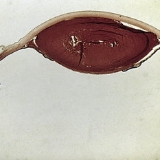
Dec 10 2012 by Yale L. Fisher, MD
This is a dislocated lens. You can see a large ovoid object resting against the ocular wall shadowing the orbital fat. Internal reflectivity demonstrates a nucleus within the larger ovoid structure. Moderate reflections from the subcapsular space and nuclear area are visible, conistent with hypermature cataract (Morgagnian type structure).
Condition/keywords: video
-
 The Effects of Blunt Trauma
Feb 27 2022 by Jesus Lozano, MD
Axial Head Ct. 60 year old man with a history of blunt trauma and lost of vision after the event. VA HM. Iop 25mmhg. Cornea clear. Complete hyphema. BMode US: diffuse Vitreous Hemorrhage with a Dislocated Lens. Retina attached.
Photographer: Dr. Jesus Lozano. Retina Specialist. Hillel Yaffe Medical Center,Israel.
Imaging device: Axial Head CT
Condition/keywords: blunt trauma, hyphema, lens dislocation
-
 Dislocated Brown Cataract with a Chorioretinal Coloboma
Sep 8 2021 by Ram Sudarshan
A 44 year-old male with dislocated brown cataract along with a chorioretinal coloboma.
Photographer: Dr.Sivadarshan
Condition/keywords: Brown cataract, chorioretinal coloboma, d, dislocated lens
 Loading…