Search results (50 results)
-
 Commotio Retinae
Mar 2 2014 by Homayoun Tabandeh, MD, FASRS
Commotio retinae following blunt trauma.
Condition/keywords: commotio retinae
-
 Commotio retinae
Jan 11 2013 by Alex P. Hunyor, MD
Commotio retinae in the temporal mid periphery in an eye which sustained blunt trauma.
Condition/keywords: Berlin's edema, commotio retinae
-
 Commotio retinae
Apr 29 2022 by Otakar Dušek, M.D. Ph.D.
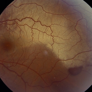
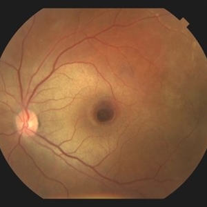
Color fundus photograph of a 24-year-old woman who was hit by a volleyball in her right eye. This caused whitening of the lower peripheral retina (Berlin's edema) i.e. commotio retinae.
Photographer: Otakar Dušek, Charles University, Prague
Imaging device: Zeiss Clarus
Condition/keywords: Berlin's edema, blunt trauma, commotio retinae
-
---thumb.jpg/image-square;max$300,300.ImageHandler) Commotio Retinae
Commotio Retinae
Jan 1 2013 by John T. Thompson, MD
commotio retinae after bullet blast near eye (non-perforating).
Condition/keywords: Berlin's edema, blunt trauma
-
 Commotio Retinae
May 19 2014 by John W. Kitchens, MD
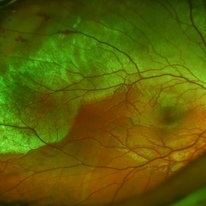
Young man with trauma.
Photographer: Ed Slade
Imaging device: Optos 200Tx
Condition/keywords: commotio retinae, trauma
-
 Commotio Retinae
May 19 2014 by John W. Kitchens, MD
Young man with trauma.
Photographer: Ed Slade
Imaging device: Optos 200Tx
Condition/keywords: commotio retinae, trauma
-
 Commotio Retinae with Retinal Hemorrhages
Mar 27 2018 by Nichole Lewis
14-year-old male hit in the right eye with a stick. Commotio Retinae with retinal hemorrhages and peripapillary hemorrhage.
Photographer: Nichole Lewis
Condition/keywords: commotio retinae, peripapillary hemorrhage, retinal hemorrhage
-
---thumb.jpg/image-square;max$300,300.ImageHandler) Severe Commotio Retinae With Edema And Hemorrhages
Severe Commotio Retinae With Edema And Hemorrhages
Dec 5 2013 by Maurice F. Rabb
Severe commotio retinae with edema and hemorrhages.
Condition/keywords: commotio retinae, edema
-
 Penetrating ocular injury with posterior impact
Dec 19 2012 by Eric A. Postel, MD
Color fundus photo of a young make s/p penetrating injury with posterior segment impact and associated hemorrhage and commotio retinae
Condition/keywords: Berlin's edema, penetration, subretinal hemorrhage
-
 Commotio Retinae
Jan 20 2015 by Andree Henaine-Berra, MD
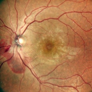
Fundus photograph of a 37-year-old male with commotio retinae and macular folds secondary to a blunt trauma due to a firecracker explosion.
Photographer: Jorge Morales-Martinez MD
Condition/keywords: blunt trauma, commotio retinae
-
 Retinal Pigment Changes After Blunt Ocular Trauma
Jun 27 2016 by Rita Couceiro, MD, MS
A 19-year-old man suffered blunt trauma of the left eye with a ball during soccer practice. At day 3 after trauma (upper pictures) the retinal area superior to the fovea looked pale and visual acuity was reduced to 20/32. This area revealed hypersignaling of retinal layers on OCT and the foveal area showed a localized disruption of retinal layers above the RPE. At day 30 (lower pictures) the retinal area of pallor showed pigmentary changes and OCT revealed atrophy of the external retinal layers. However the localized subfoveal retinal disruption was improved and only a slight disruption was seen on OCT at the ellipsoid level. Visual acuity of the left eye was restored to 20/20 although a scotoma remained.
Photographer: Rita Couceiro, Serviço de Oftalmologia do Hospital de Santa Maria, Lisboa, Portugal
Condition/keywords: blunt trauma, commotio retinae, pigment changes
-
---thumb.jpg/image-square;max$300,300.ImageHandler) Severe Commotio Retinae With Edema And Hemorrhages
Severe Commotio Retinae With Edema And Hemorrhages
Dec 5 2013 by Maurice F. Rabb
Severe commotio retinae with edema and hemorrhages.
Condition/keywords: commotio retinae, edema
-
---thumb.jpg/image-square;max$300,300.ImageHandler) Severe Commotio Retinae With Edema And Hemorrhages
Severe Commotio Retinae With Edema And Hemorrhages
Dec 5 2013 by Maurice F. Rabb
Severe commotio retinae with edema and hemorrhages.
Condition/keywords: commotio retinae, edema
-
 Chorioretinitis Sclopetaria
Jan 22 2016 by Jorge Morales-Martínez, MD
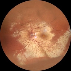
Fundus photograph of a 27-year-old male that sustained a traumatic injury in his left eye with a paintball projectile. Fundus examination showed a large subretinal hemorrhage, areas of commotio retinae and maculopathy.
Photographer: Jorge Morales-Martínez MD
-
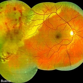 Commotio Post-Blunt Trauma
Apr 3 2018 by Paulo Bueno
Fundus photograph of an 35-year-old man with commotio retinae after indoor soccer ball blunt trauma.
Photographer: Paulo Bueno, Taubaté, Brazil.
Imaging device: Zeiss Visucam
Condition/keywords: blunt trauma, commotio retinae
-
 Commotio Retinae
Jan 20 2015 by Andree Henaine-Berra, MD
Fundus photograph of a 37-year-old male with commotio retinae and macular folds secondary to a blunt trauma due to a firecracker explosion.
Photographer: Jorge Morales-Martinez MD
Condition/keywords: blunt trauma, commotio retinae
-
 Intraocular Foreign Body
Feb 7 2019 by Somnath Chakraborty, MD
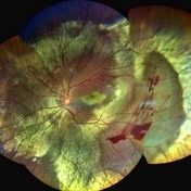
Left eye fundus photo montage of a 45-year-old male showing a large iron foreign body, impacted inferior to the infero-temporal branch vessels with a large patch of surrounding chorio-retinal atrophy, secondary to resolving Commotio retinae
Photographer: Saptarshi Mehta
Condition/keywords: commotio retinae, intraocular foreign body, trauma
-
 Chorioretinitis Sclopetaria
Jan 22 2016 by Jorge Morales-Martínez, MD
Fundus photograph of a 27-year-old male that sustained a traumatic injury in his left eye with a paintball projectile. Fundus examination showed a large subretinal hemorrhage, areas of commotio retinae and maculopathy.
Photographer: Jorge Morales-Martínez, MD
Condition/keywords: chorioretinitis sclopetaria
-
 Commotio-Retinae
Sep 22 2021 by Luiz Guilherme Freitas, MD, MsC, PhD
Fundus photograph of a 30-year-old male patient with blunt injury to the globe. Commotio retinae is retinal whitening/opacification that results from a blunt injury. The ocular findings will often resolve in a matter of days to weeks. Vision loss can result from commotio involving the posterior pole (historically referred to as Berlin’s edema). Clinical findings of commotio include the characteristic retinal whitening. Commotio may result in significant vision loss that can be transient. Healing can result in pigmentary changes and retinal thinning which may be associated with poor visual recovery if the area of involvement is macular.
Photographer: Diogo Melo, Santa Luzia Eye Hospital Recife - PE – Brazil
Condition/keywords: Berlin's edema, blunt trauma, commotio retinae, retinal whitening
-
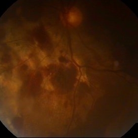
 Commotio Retinae
Apr 8 2019 by Gary R. Cook, MD, FACS
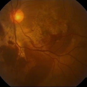
32-year-old Asian male with peripapillary and macular hemorrhages, retinal edema, and 2 concentric choroidal ruptures secondary to blunt trauma OS; V.A. = count fingers at 1 ft.
Imaging device: Topcon VT-50
Condition/keywords: blunt trauma, choroidal rupture, commotio retinae, macular hemorrhage, retinal edema
-
 A Motor Vehicle Accident Causing Valsalva Retinopathy OD, While Racing A Side By Side 4 Wheel Off-Road Vehicle
Apr 29 2020 by John S. King, MD
43-year-old white male who was injured while racing a side by side 4-wheel off-road vehicle (see Video: https://imagebank.asrs.org/file/53854/sxs-crash-during-a-race-causing-valsalva-retinopathy-od). He presented about three weeks after the injury. He was being seen by his local eye doctor who wanted an evaluation for the retinal heme and scotoma. His main complaint was a central/parcentral scotoma described as a greyish area in vision. Va 20/50 OD, nomotensive, no APD (by technician), anterior segment u/r; see picture for the fundus exam - of note there are superficial/preretinal heme, with layering of the heme superiorly, and small superficial heme at nasal edge of the optic disc; in the parafoveal region nasally there is some mottling of the RPE that may indicate an area of prior commotio retinae (also possible to have TON), which may account for his scotoma. Really bad accident (video), and amazingly, he had no LOC or injuries other than the right retina. Helmet and racing harness seat belt were used.
Photographer: Asli Ahmed
Imaging device: Topcon 50
Condition/keywords: valsalva retinopathy
-
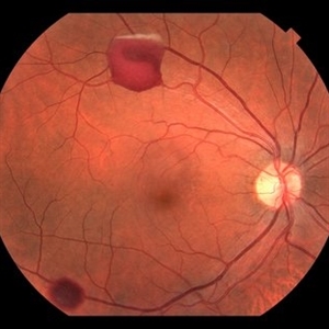
 Traumatic Pseudohole with Commotio Retinae and Subfoveal Hemorrhage after Blunt Injury
Jun 1 2019 by John S. King, MD
39-year-old African American female with central scotoma two days since blunt head injury in MVA, sent for evaluation of macular hole. 20/150 OS with IOP 12 and no RAPD. No macular hole present. Macular findings include commotio retinae and subfoveal hemorrhage.
Photographer: Stacey Coleman
Imaging device: Topcon
Condition/keywords: blunt trauma, commotio retinae, macular pseudohole, subretinal hemorrhage
-
 BERLIN'S EDEMA
Nov 21 2022 by Akansha Sharma
COLOUR FUNDUS PHOTOGRAPH OF A 35 YEAR OLD MALE WITH BERLIN'S EDEMA STATUS POST FIRE-CRACKER INJURY
Photographer: Dr. Akansha Sharma-Retina Foundation, Ahmedabad
Condition/keywords: Berlin's edema, commotio retinae, firework injury
-
 Berlin's edema
Sep 21 2023 by Vaidehi Sathaye
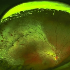
Widefield photograph of LE of a 28 year male with Berlin's edema, status post blunt trauma with tennis ball
Photographer: Dr. Vaidehi Sathaye
Imaging device: Mirante
Condition/keywords: Berlin's edema, commotio retinae
-
 Traumatic Retinal Tear
Dec 5 2021 by Aditya S Kelkar, MS, FRCS, FASRS,FRCOphth
Color fundus photograph of a 34-year old man's left eye, 2 hours after a tennis ball injury, showing commotio retinae with Berlin's edema and cherry red spot in the fovea along with linear retinal tears in the temporal equatorial zone.
Photographer: Dr Sukanya Mondal. National Institute of Ophthalmology, Pune. India.
Imaging device: Zeiss Clarus 500
Condition/keywords: Berlin's edema, cherry red spot, commotio retinae, retinal tear
 Loading…