Search results (44 results)
-
 CRAO with Cherry Red Spot
Oct 1 2012 by Jeffrey G. Gross, MD, FASRS
CRAO with cherry red spot.
Condition/keywords: central retinal artery occlusion (CRAO), cherry red spot
-
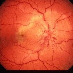
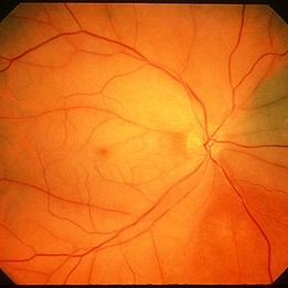
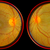
 CRAO with Cherry Red Spot and Partial Cilioretinal Artery Sparing
Oct 1 2012 by Jeffrey G. Gross, MD, FASRS
CRAO with cherry red spot and partial cilioretinal artery sparing.
Condition/keywords: central retinal artery occlusion (CRAO), cherry red spot, cilioretinal sparing
-
 CRAO with Cherry Red Spot
Oct 1 2012 by Jeffrey G. Gross, MD, FASRS
CRAO with cherry red spot.
Condition/keywords: central retinal artery occlusion (CRAO), cherry red spot
-
 AION With Ciliotretinal Artery Occlusion
May 2 2013 by Henry J. Kaplan, MD
AION accompanied by partial CRAO which is visible as retinal edema and cherry red spot.
Condition/keywords: anterior ischemic optic neuropathy, central retinal artery occlusion (CRAO)
-
 CRAO with Cherry Red Spot
Oct 1 2012 by Jeffrey G. Gross, MD, FASRS
CRAO with cherry red spot.
Condition/keywords: cherry red spot
-
 CRAO
CRAO
Mar 29 2013 by Henry J. Kaplan, MD
Central retinal artery occlusion with a cherry red spot in the fovea; notice the disc pallor.
Condition/keywords: central retinal artery occlusion (CRAO), cherry red spot
-
 Central Retinal Artery Occlusion & Cilioretinal Artery Sparing
Dec 22 2012 by Hamid Ahmadieh, MD
Color fundus photograph of the right eye of a 34-year-old man with sudden drop of vision due to CRAO. Please notice cherry red spot despite cilioretinal artery sparing .
Photographer: Zohre Salimi; Labbafinejad Medical Center, Shahid Beheshti University of Medical Sciences, Tehran
Imaging device: Topcon Fundus Camera
Condition/keywords: central retinal artery occlusion (CRAO), cherry red spot, cilioretinal sparing
-
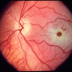
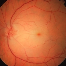
 Central Retinal Artery Occlusion
Aug 23 2012 by Gerardo Garcia-Aguirre, MD
Fundus photograph of a left eye with central retinal artery occlusion. Note the paleness of the retina (except for a very small area adjacent to the optic disc, probably irrigated by a very small cillioretinal artery), and the cherry red spot. Visual acuity is light perception.
Photographer: Noemí Hernández, Asociación para Evitar la Ceguera en México
Condition/keywords: central retinal artery occlusion (CRAO), cherry red spot, cilioretinal artery occlusion
-
 Cherry Red Spot
Mar 1 2014 by Homayoun Tabandeh, MD, FASRS
Cherry red spot in a patient with central retinal artery occlusion.
Condition/keywords: central retinal artery occlusion (CRAO), cherry red spot
-
 Central Retinal Artery Occlusion
Oct 20 2012 by Hyung-Woo Kwak, MD
This is a typical recent central retinal artery occlusion with a ‘cherry red’ spot at the macula. The patient visited our hospital with sudden visual loss occurred after the filler injection around the eyes.
Condition/keywords: central retinal artery occlusion (CRAO), cherry red spot
-
---thumb.JPG/image-square;max$300,300.ImageHandler) Central retinal artery occlusion
Central retinal artery occlusion
Oct 26 2012 by Mallika Goyal, MD
Fundus photograph of a 55-year-old gentleman one day after sudden vision loss. Shows retinal infarct with "cherry red" spot at macular centre.
Condition/keywords: central retinal artery occlusion (CRAO), cherry red spot
-
 Color Fundus Photograph of Macular Infarction Secondary to Subonjunctival Gentamicin Injection
May 16 2014 by Arwa Azmeh, MD, PhD
A 20-year-old male suffered from diplopia since age one. He was diagnosed to have acquired fourth nerve palsy in his left eye. VA at time of diagnosis was 20/20 in OU and Fundus exam was WNL in OU. His history revealed no other complaints. 3 days ago he underwent left superior oblique tucking for relief of his diplopia.The surgery was uneventful and at the end of surgery subconjunctival gentamicin was injected. Immediately following surgery his VA in OS decreased from 20/20 to complete loss of central vision and sensation of HM from the periphery. He was referred to us 3 days after surgery. At time of referral fundus exam of his left eye revealed macular infarction with cherry red spot appearance with few retinal hemorrhages, mild optic disc edema and CWS surrounding optic disc. Peripheral retina had normal color and appearance. The vitreous was clear. Anterior segment was quiet. IOP was WNL. Macular OCT was consistent with macular infarction. FA revealed delay in central retinal artery filling as fluorescein started to appear in the arteries at the level of the optic disc at 28 sec, and in the retinal veins at 38 sec. Macular area remained to be non-perfused throughout the whole FA. In late phases staining of blood vessels walls was noticed. The "wipe out" of large vessels and capillaries persisted in the central area. OCT through foveal area showed diffuse thickening of the retina with severe elevation in the fovea, reduced backscattering from the outer layers of the retina and enhanced reflectivity from the inner retina, due to ischemia. Complete blood count and cardiovascular study were WNL. The final diagnosis was macular infarction secondary to subconjunctival gentamicin injection.
Imaging device: OCT
Condition/keywords: macular infarction, subconjunctival gentamicin
-
---thumb.jpg/image-square;max$300,300.ImageHandler) Central Retinal Artery Occlusion with Cherry Red Spot
Central Retinal Artery Occlusion with Cherry Red Spot
Jun 6 2013 by Sharon Fekrat, MD FACS FASRS
Fundus photograph of a central retinal artery occlusion in the left eye. Note the cherry red spot.
Photographer: Duke Eye Imaging, Duke Eye Center, Durham, NC
Condition/keywords: central retinal artery occlusion (CRAO)
-
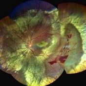
 Multiple Acute Embolic Branch Retinal Artery Occlusions
May 27 2015 by Darin R. Goldman, MD
60-year-old phakic male with multiple retinal emboli found to have a patent foramen ovale, which was repaired surgically with no further retinal occlusive episodes.
Condition/keywords: branch retinal artery occlusion (BRAO), cherry red spot, embolus, retinal microembolism
-
 CRAO
CRAO
Mar 29 2013 by Henry J. Kaplan, MD
CRAO with arterial narrowing, disc pallor,retinal edema, cherry red spot and plaques in the inferonasal artery; notice the choroidal nevus in superonasal retina.
Condition/keywords: central retinal artery occlusion (CRAO), cherry red spot
-
 OCT Through Foveal Area in Macular Infarction Secondary to Subconjunctival Gentamicin Injection
May 16 2014 by Arwa Azmeh, MD, PhD
A 20-year-old male suffered from diplopia since age one. He was diagnosed to have acquired fourth nerve palsy in his left eye. VA at time of diagnosis was 20/20 in OU and fundus exam was WNL in OU. His history reaveled no other complaints. 3 days ago he underwent left superior oblique tucking for relief of his diplopia.The surgery was uneventful and at the end of surgery subconjunctival gentamicin was injected. Immediately following surgery his VA in OS decreased from 20/20 to complete loss of central vision and sensation of HM from the periphery. He was referred to us 3 days after surgery. At time of referral fundus exam of his left eye revealed macular infarction with cherry red spot appearance with few retinal hemorrhages , mild optic disc edema and CWS surrounding optic disc. Peripheral retina had normal color and appearance. The vitreous was clear. Anterior segment was quiet. IOP was WNL. Macular OCT was consistent with macular infarction. FA revealed delay in central retinal artery filling as fluorescein started to appear in the arteries at the level of the optic disc at 28 sec, and in the retinal veins at 38 sec. Macular area remained to be non-perfused throughout the whole FA. In late phases staining of blood vessels walls was noticed. The "wipe out" of large vessels and capillaries persisted in the central area. OCT through foveal area showed diffuse thickening of the retina with severe elevation in the fovea, reduced backscattering from the outer layers of the retina and enhanced reflectivity from the inner retina, due to ischemia. Complete blood count and cardiovascular study were WNL. The final diagnosis was macular infarction secondary to subconjunctival gentamicin injection.
Imaging device: OCT
Condition/keywords: macular infarction, subconjunctival gentamicin
-
 Late FA Phase of Macular Infarction Secondary to Subconjunctival Gentamicin Injection
May 16 2014 by Arwa Azmeh, MD, PhD
A 20-year-old male suffered from diplopia since age one. He was diagnosed to have acquired fourth nerve palsy in his left eye. VA at time of diagnosis was 20/20 in OU and fundus exam was WNL in OU. His history revealed no other complaints. 3 days ago he underwent left superior oblique tucking for relief of his diplopia.The surgery was uneventful and at the end of surgery subconjunctival gentamicin was injected. Immediately following surgery his VA in OS decreased from 20/20 to complete loss of central vision and sensation of HM from the periphery. He was referred to us 3 days after surgery. At time of referral fundus exam of his left eye revealed macular infarction with cherry red spot appearance with few retinal hemorrhages, mild optic disc edema and CWS surrounding optic disc. Peripheral retina had normal color and appearance. The vitreous was clear. Anterior segment was quiet. IOP was WNL. Macular OCT was consistent with macular infarction. FA revealed delay in central retinal artery filling as fluorescein started to appear in the arteries at the level of the optic disc at 28 sec, and in the retinal veins at 38 sec. Macular area remained to be non-perfused throughout the whole FA. In late phases staining of blood vessels walls was noticed. The "wipe out" of large vessels and capillaries persisted in the central area. OCT through foveal area showed diffuse thickening of the retina with severe elevation in the fovea, reduced backscattering from the outer layers of the retina and enhanced reflectivity from the inner retina, due to ischemia. Complete blood count and cardiovascular study were WNL. The final diagnosis was macular infarction secondary to subconjunctival gentamycin injection.
Imaging device: OCT
Condition/keywords: macular infarction, subconjunctival gentamicin
-
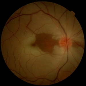
 Central Retinal Artery Occlusion With Cilioretinal Sparing
Apr 4 2018 by Soumya Venkatesh
Fundus photograph of a 23-year-old gentleman presenting with sudden loss of vision 2 days prior to presentation. He underwent all relevant investigations and found to have APLA positive. He also had dengue serology positive. On follow up, his retinal edema reduced unmasking the underlying hemorrhages( flame shaped).
Photographer: Soumya Harapanahalli Venkatesh, JSS university, Karnataka, India
Condition/keywords: central retinal artery occlusion (CRAO), cherry red spot, cilioretinal sparing, retinal ischemia
-
 Early-FA-phase-of-macular-infarction-secondary-to-subconjunctival-gentamycin-injection
May 16 2014 by Arwa Azmeh, MD, PhD
A 20-year-old male suffered from diplopia since age one. He was diagnosed to have acquired fourth nerve palsy in his left eye. VA at time of diagnosis was 20/20 in OU and Fundus exam was WNL in OU. His history revealed no other complaints. 3 days ago he underwent left superior oblique tucking for relief of his diplopia.The surgery was uneventful and at the end of surgery subconjunctival gentamicin was injected. Immediately following surgery his VA in OS decreased from 20/20 to complete loss of central vision and sensation of HM from the periphery. He was referred to us 3 days after surgery. At time of referral fundus exam of his left eye revealed macular infarction with cherry red spot appearance with few retinal hemorrhages, mild optic disc edema and CWS surrounding optic disc. Peripheral retina had normal color and appearance. The vitreous was clear. Anterior segment was quiet. IOP was WNL. Macular OCT was consistent with macular infarction. FA revealed delay in central retinal artery filling as fluorescein started to appear in the arteries at the level of the optic disc at 28 sec, and in the retinal veins at 38 sec. Macular area remained to be non-perfused throughout the whole FA. In late phases staining of blood vessels walls was noticed. The "wipe out" of large vessels and capillaries persisted in the central area. OCT through foveal area showed diffuse thickening of the retina with severe elevation in the fovea, reduced backscattering from the outer layers of the retina and enhanced reflectivity from the inner retina, due to ischemia. Complete blood count and cardiovascular study were WNL. The final diagnosis was macular infarction secondary to subconjunctival gentamicin injection.
Imaging device: OCT
Condition/keywords: macular infarction, subconjunctival gentamicin
-
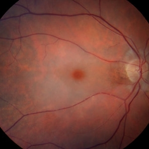
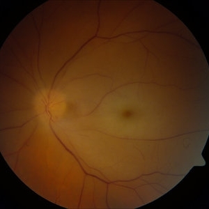 Central Retinal Artery Occlusion
Feb 28 2013 by Theodore Leng, MD, MS, FASRS
Color fundus photograph of a 64-year-old man with a central retinal artery occlusion. Note the cherry red spot.
Imaging device: Zeiss FF450
Condition/keywords: central retinal artery occlusion (CRAO)
-
 Central Retinal Artery Occlusion
Jun 4 2019 by Unnati Vishwanath Shukla, M. S. ,DNB, FVRS FNERF, MNAMS,PhD Scholar(Retina)
A young female patient of Indian origin on Oral Contraceptive medication presenting with Central Retinal Artery Occlusion with Cilioretinal artery Sparing.
Photographer: Unnati Shukla, C.H. Nagri Eye Hospital, NHL medical college, Ahmedabad,Gujarat,India.
Condition/keywords: central retinal artery occlusion (CRAO), cherry red spot, cilioretinal sparing, pale retina
-
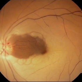
 Traumatic Retinal Tear
Dec 5 2021 by Aditya S Kelkar, MS, FRCS, FASRS,FRCOphth
Color fundus photograph of a 34-year old man's left eye, 2 hours after a tennis ball injury, showing commotio retinae with Berlin's edema and cherry red spot in the fovea along with linear retinal tears in the temporal equatorial zone.
Photographer: Dr Sukanya Mondal. National Institute of Ophthalmology, Pune. India.
Imaging device: Zeiss Clarus 500
Condition/keywords: Berlin's edema, cherry red spot, commotio retinae, retinal tear
-
 Central Retinal Artery Occlusion
Jan 13 2020 by Prithvi Chandrakanth
37-years-old male with complaints of sudden diminution of vision in the left eye for the past three days. Fundus examination revealed pale retina in the left eye with cherry red spot and normal fundus picture in right eye.
Photographer: DR.PRITHVI CHANDRAKANTH, ARAVIND EYE HOSPITAL, UDUMALPET
Imaging device: TRASH TO TREASURE RETCAM
Condition/keywords: central retinal artery occlusion (CRAO), cherry red spot, retcam, smartphone fundus photography
-
 BRAO with Cherry Red Spot
Mar 28 2020 by Sugnesh Parmar
20-year-old male presented with sudden loss of vision in his RE and on examination found to have BRAO with typical cherry red spot.
Photographer: Dr. Sugnesh Parmar, Radheshyam retina hospital, Bhavnagar, Gujarat, India
Condition/keywords: branch retinal artery occlusion (BRAO)
-
 Central Retinal Artery Occlusion
Mar 26 2019 by Gary R. Cook, MD, FACS
61-year-old male patient with acute CRAO OS demonstrating a hyperemic optic disc with a couple of peripapillary hemorrhages, generalized arteriolar narrowing, a cherry-red spot in the macula, and retinal whitening surrounding the fovea; VA= LP.
Imaging device: Topcon VT-50
Condition/keywords: central retinal artery occlusion (CRAO), cherry red spot, retinal whitening
 Loading…