Search results (321 results)
-
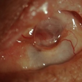
 "Flower Cataract"
Jul 11 2013 by Jason S. Calhoun
Patient presents with a cataract shaped like a flower. Patient had surgery to remove cataract.
Photographer: Jason S. Calhoun, Department of Ophthalmology, Mayo Clinic Jacksonville, Florida
Condition/keywords: cataract
-
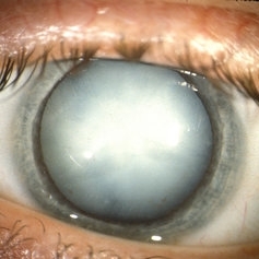 Hypermature Cataract
Oct 8 2012 by Jeffrey G. Gross, MD, FASRS
Hypermature cataract HM.
Condition/keywords: hypermature cataract
-
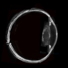
 Ciliary Body Melanoma With Partial Ring Configuration and Diffuse Sentinel Vessels
Feb 26 2014 by Susanna S. Park, MD, PhD
Slit lamp photo of a 57-year-old man with new vision loss from cataract formation. Large ciliary body mass with diffuse sentinel vessels is noted. The eye was removed and the tumor was noted to have a partial ring configuration with predominantly epithelioid cells and early vitreous seeding.
Photographer: Ellen Redenbo, University of California Davis Eye Center
Condition/keywords: ciliary body melanoma, melanoma
-
 Whole Eye OCT
Jan 4 2019 by Netan Choudhry, MD, FRCS(C) FASRS
Swept-Source OCT montage of a 45-year-old male with Alports disease and posterior subcapsular cataract.
Photographer: John Golding BA, Vitreous Retina Macula Specialists of Toronto
Imaging device: Topcon DRI Triton
Condition/keywords: Alports disease, optical coherence tomography (OCT), swept source
-
---thumb.JPG/image-square;max$300,300.ImageHandler) TID (Trans Illumination Defect)
TID (Trans Illumination Defect)
Jul 8 2013 by Jason S. Calhoun
74-year-old patient who VA 20/70 OD, 20/50 OS. Complaints of blurred vision. Iris atrophy, both eyes. ERM, right eye, Patient to have cataract surgery to improve distance vision.
Photographer: Jason S. Calhoun, Department of Ophthalmology, Mayo Clinic Jacksonville, Florida
Condition/keywords: translucency of iris
-
 Elschnig's Pearls
Sep 1 2015 by René Hernán Parada Vásquez
Fundus photograph of 58-year-old male with Elschnig's pearls, you can see the transparent clusters formed by proliferation of epithelial lens cells found in the remains of the capsule of the crystalline lens following cataract surgery.
Photographer: Parada René, ESO, Guatemala.
Condition/keywords: cataract surgery
-
 "Flower Cataract"
Jul 11 2013 by Jason S. Calhoun
Patient presents with a cataract shaped like a flower. Patient had surgery to remove cataract.
Photographer: Jason S. Calhoun, Department of Ophthalmology, Mayo Clinic Jacksonville, Florida
Condition/keywords: cataract
-
 Rubella cataract
May 2 2013 by Henry J. Kaplan, MD
Cataract due to rubella infection; #1.
Condition/keywords: cataract, rubella retinopathy
-
 Closed Funnel Retinal Detachment
Apr 9 2017 by Aliya Sultana
Fundus phtograph of an 51-year-old man with closed funnel rhegmatogenous retinal detachment presented to our department 6 weeks after cataract surgery. Posterior capsule rent noticed with vitreous in anterior chamber, condensed vitreous tag is incarcerated in side port wound.
Photographer: Dr Aliya Sultana , Assistant Professor,Sarojini Devi Eye Hospital, Hyderabad, Telangana. India.
-
 Superior Peripapillary Hemorrhage
Jul 13 2013 by Jason S. Calhoun
Patient was seen for acute vision loss in the right eye. Patient has glaucoma. VA was 20/70 in the right eye. Had vitrectomy back in May 2012 for ERM stripping. Also had trabectome with cataract surgery in December of 2012. Fundus photos presents a superior peripapillary Hemorrhage of the optic nerve. Patient will be followed up in one month.
Photographer: Jason S. Calhoun, Department of Ophthalmology, Mayo Clinic Jacksonville, Florida
Imaging device: TOPCON TRC 50-EX
Condition/keywords: peripapillary hemorrhage
-
 Vitreous Hemorrhage
Nov 9 2012 by Norman Byer
This 60-year-old man suddenly developed a vitreous hemorrhage from this acute horseshoe tear 3½ years following cataract extraction when a posterior vitreous detachment occurred. The white nubbin identifies this lesion as a preexisting cystic retinal tuft. The pigment spot beneath the flap is evidence of secondary trophic changes in the pigment epithelium. Note the irregular shape of the flap with the narrow tip and broad base. This was caused by vitreous traction which was exerted at two separate points on the retina and which tore the retina at each place.
Condition/keywords: acute posterior vitreous detachment, irregularly shaped flap, trophic pigmented changes, vitreous hemorrhage, vitreous traction, white retinal tuft
-
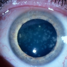
---thumb.jpg/image-square;max$300,300.ImageHandler) Retinitis Pigmentosa and Cataract
Retinitis Pigmentosa and Cataract
Dec 27 2013 by David Callanan, MD
23-year-old male patient, 20/100 OU.
Condition/keywords: cataract, retinitis pigmentosa
-
---thumb.jpg/image-square;max$300,300.ImageHandler) Diabetes with Cortical Cataract
Diabetes with Cortical Cataract
Apr 4 2014 by H. Michael Lambert, MD
Diabetes. Cortical Spoking
Photographer: Donald Lowd
Condition/keywords: cataract, diabetes
-
000---thumb.jpg/image-square;max$300,300.ImageHandler) Dropped IOL into the Vitreous Cavity
Dropped IOL into the Vitreous Cavity
Oct 7 2012 by Young Hee Yoon, MD, PhD
Fundus photograph of an 70-year-old man with a history of cataract operation 20 years ago. He visited our clinic with decreased visual acuity for 2 days.
Photographer: Yoon-hwa Kim, Asan Medical Center
Imaging device: Optomap, optos
Condition/keywords: intraocular lens dislocation
-
---thumb.JPG/image-square;max$300,300.ImageHandler) Polychromatic Cataract (Christmas Tree Cataract)
Polychromatic Cataract (Christmas Tree Cataract)
Jul 8 2013 by Jason S. Calhoun
Patient came in for cataract evaluation with blurred vision in the left eye. Patient will think about surgery and call back to schedule.
Photographer: Jason S. Calhoun, Department of Ophthalmology, Mayo Clinic Jacksonville, Florida
Condition/keywords: cataract
-
---thumb.jpg/image-square;max$300,300.ImageHandler) Congenital Cataracts
Congenital Cataracts
Dec 26 2013 by David Callanan, MD
42-year-old female patient, 20/30 OU; high myope.
Condition/keywords: congenital cataract, high myopia
-
 Traumatic Lens Drop in Vitreous
Dec 15 2020 by Manish Nagpal, MD, FRCS (UK), FASRS
Patient had come to us status post blunt trauma with the lens dislocated in inferior vitreous.
Photographer: Gayathri Mohan, Retina Fellow, Retina Foundation, Ahmedabad, India
Imaging device: Mirante CSLO
Condition/keywords: dropped nucleus, lens dislocation, traumatic cataract
-
---thumb.JPG/image-square;max$300,300.ImageHandler) Polychromatic Cataract (Christmas Tree Cataract)
Polychromatic Cataract (Christmas Tree Cataract)
Jul 8 2013 by Jason S. Calhoun
Patient came in for cataract evaluation with blurred vision in the left eye. Patient will think about surgery and call back to schedule.
Photographer: Jason S. Calhoun, Department of Ophthalmology, Mayo Clinic Jacksonville, Florida
Condition/keywords: cataract
-
 Bergmeister's Papilla
Sep 29 2020 by Dhaivat Shah
Bergmeister's papilla is a small tuft of glial tissue which arises from the center of the optic disc, and represents a remnant of the fetal hyaloid artery. The hyaloid artery provides nutrition to the lens during development, and runs forward to the lens from the optic disc. At birth the hyaloid artery regresses, and is normally completely regressed by the time of birth. Bergmeister's papilla is frequently observed as an incidental clinical finding if this artery has an incomplete regression posteriorly. However, in the severe forms it can be associated with cataracts, persistence of the primitive vitreous, microphthalmia, vitreous hemorrhages and sometimes tractional retinal detachment, due to contraction of the residual fibro vascular tissue. Therefore, careful monitoring of vitreous thickening in the peripapillary areas, both by examining the ocular fundus, and especially by SD-OCT, is of considerable importance. Here we have one such of a 30 year old young male who came in for a routine checkup, in whom we noted a Bergmeister’s papilla. Due to its benign nature, patient was reassured and was asked to follow up yearly.
Condition/keywords: Bergmeister's Papillae
-
 Intra Lenticular Foreign Body
Jan 1 2013 by John T. Thompson, MD
Trauma to equatorial lens with small retained foreign body within lens, lens remained clear.
Condition/keywords: intralenticular foreign body, traumatic cataract
-
---thumb.JPG/image-square;max$300,300.ImageHandler) Polychromatic Cataract (Christmas Tree Cataract)
Polychromatic Cataract (Christmas Tree Cataract)
Jul 8 2013 by Jason S. Calhoun
Patient came in for cataract evaluation with blurred vision in the left eye. Patient will think about surgery and call back to schedule.
Photographer: Jason S. Calhoun, Department of Ophthalmology, Mayo Clinic Jacksonville, Florida
Condition/keywords: cataract
-
---thumb.jpg/image-square;max$300,300.ImageHandler) Traumatic Dislocation of Hypermature Cataract
Traumatic Dislocation of Hypermature Cataract
Jan 1 2013 by John T. Thompson, MD
Hypermature cataract with traumatic dislocation into anterior chamber.
Condition/keywords: anterior dislocation of lens, blunt trauma, hypermature cataract
-
 Panophthalmitis
Jul 12 2014 by Philip J. Polkinghorne, MD
A 85-year-old lady who presented with an eroding intraocular lens. She had been initially treated with herbal medicines which failed to control the infection.
Photographer: Philip Polkinghorne
Condition/keywords: cataract surgery, endophthalmitis, panophthalmitis
-
 24 Hours Post Scleral Wound Closure+ Scleral Buckle+25 g Vitrectomy+Silicon Oil
Jan 23 2015 by Carlos Quezada-Ruiz, MD, FASRS
24 hours post op fundus photograph of a 43-year-old man who had perforating injury to the right eye with a small piece of plastic while he was hammering. OD LP, subconjunctival hemorrhage, clear cornea, hyphema, irido and ciclodyalisis as well as a luxated lens with traumatic cataract and a dense vitreous hemorrhage. B-US showed rhegmatogenous retinal detachment with a tear and a big inferior hemorrhagic choroidal detachment. 360 peritomy revealed 2-entry scleral wounds were found in zone II (M V and M VI) and closure was performed. 25 G PPV was performed with the infusion canal placed in the AC through the limbus. Lensectomy and removal of a dense recent vitreous hemorrhage revealed a white detached retina with an exit wound through the temporal inferior segment of the optic nerve with a nasal GRT and sub retinal hemorrhage as well as temporal inferior choroidal, PVD was induced and PFOs helped stabilizing the retina while vitrectomy and sub-retinal hemorrhage was removed through the GRT. Fluid air exchange was made and 360 endolaser over the buckle indentation was done and silicon oil was used as endotamponade. This picture was taken 24 hrs after the surgery.
Photographer: Lilibeth Rodriguez, Instituto de la Visión. Torreon, Mexico.
Condition/keywords: central retinal artery occlusion (CRAO), giant retinal tear, trauma
-
 Superior Peripapillary Hemorrhage
Jun 27 2013 by Jason S. Calhoun
Patient was seen for acute vision loss in the right eye. Patient has glaucoma. VA was 20/70 in the right eye. Had vitrectomy back in May 2012 for ERM stripping. Also had trabectome with cataract surgery in December of 2012. Fundus photos presents a superior peripapillary Hemorrhage of the optic nerve. Patient will be followed up in one month.
Photographer: Jason S. Calhoun, Mayo Clinic Jacksonville, Florida
Imaging device: TOPCON TRC 50-EX
Condition/keywords: peripapillary hemorrhage
 Loading…